Lymphatic System
Lymphatic System. ST120: Concorde Career College. Lymphatic System . Objectives: Define the term lymph. Describe the functions of the lymphatic system. List and identify the structures of the lymphatic system and describe the function of each. Identify the origin of lymph.

Lymphatic System
E N D
Presentation Transcript
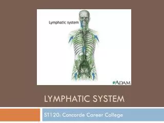
Lymphatic System ST120: Concorde Career College
Lymphatic System Objectives: • Define the term lymph. • Describe the functions of the lymphatic system. • List and identify the structures of the lymphatic system and describe the function of each. • Identify the origin of lymph.
Lymphatic System Objectives: • Trace the flow of lymph through the body. • Describe the immune system and describe the immune response. • Describe the mechanism by which the immune system helps to maintain homeostasis.
Lymphatic System Objectives: • Describe common diseases, disorders, and conditions of the lymphatic system including signs and symptoms, diagnosis, and available treatment options. • Demonstrate knowledge of medical terminology related to the lymphatic system verbally and in the written form.
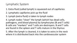
Lymphatic System Functions of the Lymphatic System • Removal of impurities • Lymphocyte processing
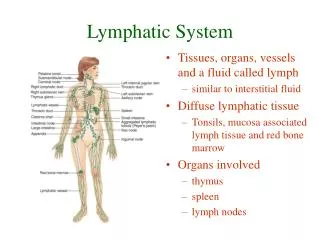
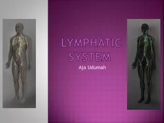
Lymphatic System • The lymphatic system parallels (same direction)the circulatory system • Lymph vessels return tissue fluid to the blood & protect the body against foreign material • The lymphatic system includes: • Lymph fluid • Lymph vessels • Lymph nodes • Spleen • Thymus • And tonsils
Lymph Fluid Lymph is the tissue fluid that enters and travels through the lymphatic vessels • Plasma flows out of blood capillaries into tissue spaces (interstitial fluid) – AKA Tissue fluid • Plasma diffuses out of the capillaries faster than it is reabsorbed, the remaining interstitial fluid is returned to the blood through the lymphatic system
Lymphatic Capillaries • Lymphatic Capillaries - Carry lymph and resemble blood capillaries • Lacteals - Specialized lymphatic capillaries that carry chyle
Lymph Vessels • Vessels of the lymphatic system start with the smallest blind (dead-ended) lymph capillaries • The walls of the lymphatic capillaries are a thin walled, single layer of loosely fit squamous epithelial cells making them highly permeable • Lymphatic capillaries join larger lymphatic arterioles that then join lymphatic arteries • Lymphatic vessels like the veins of the circulatory system have valves
Lymphatic System Relationship of Lymphatic Vessels to Circulatory System
Thoracic Duct • Lymph returns to the blood flow through the thoracic duct and the left subclavianvein • Lymph from the lower body returns through the thoracic duct • Cisterna chyli– dilation at the beginning of the thoracic duct (pouch-like structure) • Lymph from a portion of the upper body also empties into the thoracic duct superior to the cisternachyli • Lymph from the upper left quadrant empties into the subclavian vein • Two terminal vessels include: Right Lymphatic duct & thoracic duct
Lacteal • Found in the villi of the small intestine • Responsible for absorbing fat-soluble nutrients (fatty acids and vit. A,D,E & K) • Fats are absorbed by the lacteals as chylomicron • Chylomicron: combination of fatty acids and glycerol to form triglycerides
Lymph nodes and nodules • Lymph Node: small ball shaped organs of the lymphatic system • Lymph Nodules: groups of lymph cells located beneath the epithelial layer of mucus membranes (ex. Respiratory tract and digestive tract) • Peyer’s patches: lymphatic nodules of the ileum • Filters pathogens and foreign material before returning to blood • Monitor the level of body fluid • Hematopoietic function – produce lymphocytes, macrophages, and monocytes • Located throughout the body • Cervical • Axillary • Inguinal
Lymph node Hilus Afferent vessels x4 pg.261 A&P Efferent vessels x1 Capsule Germinal centers Sinuses Trabecula
Tonsils • Palatine Tonsils (throat) – located at each side of the oropharynx • Pharyngeal (Adenoids) – located on the posterior wall of the nasopharynx • Lingual Tonsils – located at the base of the tongue
Lymphedema • Abnormal accumulation of fluid in interstitial spaces • Causes: • Obstruction of a lymphatic vessel (tumors, inflammation) • Increased capillary BP • Abnormal uptake of fluid by the lymphatic capillaries due to trauma or surgical wound (often seen after mastectomy)
Terms • Lymphadenitis: inflammation of lymph node/vessels • Metastasis: is the spread of a disease from one organ or part to another non-adjacent organ or part, causing a new cancer site • Lymphadenopathy: Any disease that usually involves the lymph glands
Sentinel Lymph Node Biopsy • Method developed to determine if breast cancer has spread to lymph ducts or nodes in the axilla • Prevents the patient from having to undergo a lymph node dissection • Sentinel Node: first node the breast drains to Nuclear Medicine • A radioactive dye is injected into the area where the tumor is located as well as around the areola • Hours later photos will reveal the pathway used to exit the breast, this will reveal the sentinel node OR • Surgeon injects a blue dye into the breast that assists in visual of the nodes • Small incision is made in the axilla • A special probe picks up signals form the radioactive dye and gets “excited” when over an affected node • All blue and radioactive nodes are removed
Spleen • Highly vascular and friable (easily crumbled/delicate) • Located behind the stomach • Contains lymphoid nodules (white pulp) • Filters blood (red pulp) cleans out foreign materials/microorganisms • Stores blood (approximately 200 ml) aids in maintenance of BP during severe hemorrhage • Hematopoietic function: lymphocytes, monocytes, leukocytes, fixed plasma cells • In a fetus the spleen produces erethrocytes
Spleen Hilus Splenic artery Splenic vein
Spleen cont. • #1 organ injured in MVA’s (motor vehicle accidents) despite being protected by the lower ribs • Splenectomy: surgical removal of the Spleen
Lymphatic System Reticuloendothelial System • Responsible for destruction of obsolete RBCs, bacteria, cancer cells, and other potentially harmful foreign substances. • Utilizes monocytes distributed throughout the body such as Kupffer’s cells located in the liver
Thymus • Located in the mediastinum • Composed of two lateral lobes bound by a connective tissue capsule • Proper function dependent on thymosin • Produces T-cells important in cell mediated immunity
Lymphatic System Lymphatic and Immune Systems • Thymus • Bone marrow • Spleen • Tonsils • Lymph nodes • Lymph capillaries • Lymph vessels • Lymphocytes • Lymph
Lymphatic System Immunologic Defenses • Provide the body with general defenses against disease. • Nonspecific Defenses • Specific Defenses
Lymphatic System Nonspecific Defenses • Chemical and Mechanical Barriers • Phagocytosis • Natural Killer Cells • Inflammation • Fever • Interferon
Lymphatic System Occurrence of Infection • Not every exposure to a pathogen results in infection. • Portal of Entry • Virulence • Toxins • Dose • Predisposition
Lymphatic System Barriers Against Infection • Chemical • Body secretions • Mechanical • Skin • Mucous membranes • Cilia
First Line of Defense • Our first line of defense against microbes include: • Skin • Mucous membranes • Secretions
Skin • Unbroken skin prevents microbes from entering the body (keratin) • Broken skin such as Burns, Cuts, Scratches open the doors for an invasion of microbes such as Staphylococcus aureus • Sweat glands and Sebaceous gland secrete acids that kill or inhibit the growth of bacteria on the skin
Mucous Membranes • Line tracts open to the exterior of the body (respiratory, digestive, urinary) • Goblet cells secrete mucous to line the membranes and “catch” microbes • Cilia catch and pass debris up to the pharynx to be expelled • Glands in the stomach produce highly acidic gastric juice that kills most bacteria • Helicobacter Pylori is an exception… can cause stomach and duodenal ulcers
Second Line of Defense • If pathogens penetrate the 1st line of defense they will encounter the body’s 2nd line of defense Circulatory and chemical defenses • Circulatory Defenses: • Phagocytes • Complement proteins • Iron • Fever • Inflammatory response • Interferons
Phagocytes • Leukocytes responsible for phagocytosis • Phagocytosis: the process of surrounding and ingesting another microbe • Granulocytes • Neutrophils – motile and effective phagocytes • Eosinophils – attach to parasites and secrete peroxide ions which kill them. Play a larger phagocytic role during allergic response • Basophils • Agranulocytes • Monocytes – enter body tissues and mature into stationary macrophages or travel through blood to infection site (wandering macrophages) • Kupffer cells (liver), microglia (brain) • Lymphocytes – are not phagocytes but are important in the immune response
Phagocytosis • Chemotaxis – chemicals attract phagocytes to injured tissues or bacteria • Adherence – antibody molecules secrete a serum protein that can coat a bacterial cell and promote attachment (opsonization) • Ingestion – phagocyte surrounds the microbe • Pseudopods: “false feet” that extent to cover a microbe • The resulting sac is called a Phagosome • Digestion – phagosome is moved into the cytoplasm (phagolysosome) and enzymes (from the lysosomes) destroy the bacteria • Degranulation – digestion process
Complement proteins • Group of 25-30 serum proteins found in blood plasma • Circulate inactive until needed in the antibody-antigen reaction • Activated Proteins: increase inflammatory response, assist in lysis of antigen, attracts phagocytes • Complement cascade: sequence of proteins “bore” holes into bacteria causing an accumulation of fluid that eventually ruptures the cell
Iron • Virulence of bacteria increases when free iron is present • Leukocytes: produce interleukin-1 (IL-1), stimulates the liver to store iron depriving bacteria of necessary free iron
Inflammatory response • Inflammation is a beneficial and necessary response to injury • The four signs of inflammation include: • Pain • Heat • Redness • Swelling • Three stages of inflammation include: • Vasodilatation • Phagocyte migration • Tissue repair
Inflammatory Response • Vasodilation: Increased blood flow to area and increased permeability of blood vessels • Histamine, kinins, and prostaglandins: chemicals released by damaged tissues responsible for vasodilatation
Fever • An abnormally elevated body temperature (controlled by hypothalamus) • Systemic response frequently caused by viral or bacterial invasion • Symptoms include heat, shivering and increased metabolism rate • Vasodilatation and sweating signify the temperature falling “ fever breaking”
Interferons- produced by T-cells • Released to protect other cells by interferring with the ability of the virus to reproduce as it moves from cell to cell • Proteins produced by leukocytes, T-lymphocytes, and fibroblasts in response to infection “interfere with viral replication” • Proteins enter surrounding cells and inhibit the synthesis of proteins needed by invading viruses for multiplication • They interfere with viral replication Pg. 269 A&P