Lymphatic System
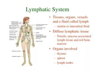
Lymphatic System. CHAPTER 22. Introduction. The lymphatic system consists of a fluid called lymph flowing within lymphatic vessels, several structures and organs that contain lymphatic tissue, and red bone marrow which houses stem cells that develop into lymphocytes.

Lymphatic System
E N D
Presentation Transcript
Lymphatic System CHAPTER 22
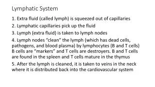
Introduction • The lymphatic system consists of a fluid called lymph flowing within lymphatic vessels, several structures and organs that contain lymphatic tissue, and red bone marrow which houses stem cells that develop into lymphocytes. • The composition of the interstitial fluid and lymph are basically same. • After fluid passes from interstitial spaces into lymphatic vessels, it is called lymph. • Lymphatic tissue is a specialized type of conn. Tissue that contains large number of lymphocytes.
Functions of the Lymphatic System • Draining interstitial fluid: lymphatic vessels drain excess interstitial fluid from tissue spaces. • Transporting dietary lipids: lymphatic vessels transport the lipids and lipid-soluble vitamins (A,D, E, K) absorbed by the GI tract to the blood. • Facilitating immune response: lymphatic tissue initiates highly specific responses directed against particular microbes or abnormal cells • lymphocytes can recognize foreign cells, microbes, toxins and cancer cells. They respond in two ways.
Functions continued • The T-lymphocytes or T-cells destroy the intruders by causing them to rupture or by releasing cytotoxic substances. • The B-cells secrete antibodies which cause destruction of specific antigens.
Lymphatic Vessels and Lymph Circulation • Lymphatic vessels begin as lymphatic capillaries. • Lymphatic capillaries are found throughout the body except in avascular tissues, the CNS, portions of the spleen and red bone marrow. • They are closed-ended tubes located in spaces between cells. • These unite to form larger lymphatic vessels. They resemble veins but have thinner walls and more valves. • At various intervals along the lymphatic vessels, lymph flows through structures called lymph nodes. • In the skin, lymphatic vessels are in the subcutaneous tissue and in the viscera follow arteries, form plexuses.
Lymphatic Capillaries • They are slightly larger than blood capillaries. • The wall is made of endothelial cells. The margins of the cells overlap. • When pressure is greater in the IF than in lymph, the cells separate and fluid enters the capillary. • When pressure is greater in the capillary the cells adhere closer together, so lymph cannot flow back to IF.
Capillaries • Anchoring filaments: attach lymphatic endothelial cells to surrounding tissues. • When excess IF accumulates the filaments are pulled, to make the openings larger so fluid can enter the capillary. • In the SI, the capillaries are called lacteals. • These carry dietary lipids into lymphatic vessels and ultimately into blood. • The presence of these lipids causes the lymph draining the SI to appear creamy white and is called chyle.
Lymph Trunks • Lymph passes form lymphatic capillaries, through lymph vessels and then lymph nodes. • The lymphatic vessels that exit nodes, pass lymph either toward another node or on to another group of nodes. • From the most p[proximal group of each chain of nodes, the exiting vessel form lymph trunks. • The principal trunks are lumbar, intestinal, bronchomediastinal, subclavian and jugular trunks. • The principal trunks pass their lymph into two min channels, thoracic and right lymphatic duct. Lymph then passes into venous blood.
Lymphatic Ducts • The thoracic (left lymphatic) duct-this is about 38-45 cm long. Begins as cisterna chyli ant to second lumbar vertebra. This receives lymph from the left side of the head, neck and chest, the left upper limb, and the entire body inferior to the ribs. This drains lymph into venous blood via the left subclavian vein. • The cisterna chyli receives lymph from the right and left lumbar trunks and from the intestinal trunk. • Lumbar trunk-from lower limbs, wall and viscera of pelvis, kidneys, adrenal glands. • Intestinal trunk: from stomach, intestines, pancreas, spleen and part of liver.
Lymphatic Ducts • In the neck the thoracic duct receives lymph from the left jugular. Left subclavian and left bronchomediastional trunks. • The right lymphatic duct is about 1.25 cm long and drains lymph from the upper right side of the body into venous blood via the right subclavian vein. • Three lymphatic trunks drain into this duct-right jugular:right side of head and neck. Right subclavian:right upper limb. Right bronchomediastinal:right sides of thorax, lung heart and part of liver.
Formation and Flow of Lymph • More fluid if filtered out of the capillaries than is reabsorbed. • This excess filtered fluid-3L a day- drains into lymphatic vessels and becomes lymph. • Function of lymphatic vessels is to return the lost plasma proteins back to blood. • Ultimately, lymph drains into venous blood through the right lymphatic and thoracic duct at the junction of internal jugular and subclavian veins.
Relationship of lymphatic system to cardiovascular system • Sequence of fluid flow is capillary (blood) ->interstitial spaces (IF)->lymphatic capillaries (lymph)->lymphatic vessels (lymph)->lymphatic ducts (lymph)->subclavian veins (blood). • The skeletal muscle and respiratory pumps promote the flow of lymph from tissue spaces to the large lymphatic ducts to the subclavian veins. • Lymphatic vessels posses one-way valves, similar to veins to prevent back flow.
Lymphatic Organs and Tissues • The organs and tissues are classified into two groups based on the functions. • Primary lymphatic organs:provide appropriate environment for stem cells to divide and mature into B cells and T cells. The organs are the red bone marrow (in flat bone and epiphysis of ling bones of adults) and thymus gland. • Secondary lymphatic organs: most immune responses occur. Include lymph nodes, spleen and lymphatic nodules.
Thymus Gland • The thymus gland lies between the sternum and the two large blood vessels above the heart. • It usually has two lobes. A connective tissue layer holds the two lobes together. A connective tissue capsule encloses each lobe separately. • Lobes are divided into lobules. • Each lobule made of an outer cortex: made of lymphocytes, epithelial cells and an inner medulla also of reticular epithelial cells. These produce thymic hormones aid in maturation of T cells. Thymic corpuscles. • Thymus gland in infants=70 g and in adults=3 g.
Spleen • The spleen is an oval shaped organ and is the largest mass of lymphatic tissue. • It is located in the left hypochondriac region between the stomach and diaphragm. • A capsule of dense connective tissue surrounds the spleen. • The parenchyma of spleen consists of two diff. Kinds of tissues-white pulp and red pulp.
Spleen • White pulp: lymphatic tissue, mostly lymphocytes and macrophages arranged around branches of the splenic artery called central arteries. Fn.-B and T cells carry out immune functions. Macrophages destroy blood-borne pathogens. • Red pulp consists of venous sinuses filled with blood and cords called splenic cords. This consists of red blood cells, macrophages, lymphocytes, plasma cells and granulocytes.Fn.-removal by macrophages of worn out or defective RBC’s and platelets. Storage of platelets and hemopoiesis during fetal life. • Abdominal trauma-splenectomy to prevent shock. Red bone marrow and liver take over its function.
Lymph Nodes • There are approx. 600 of these bean-shaped organs. • Located along lymphatic vessels. • Heavily concentrated near the mammary glands and in the axillae and groin. • Are about 1-25mm long. Covered by a capsule of dense conn. Tissue. • Capsular extensions called traberculae divide it into compartments. Provide support and a route for blood vessels t pass. • Internal to this is supporting network of reticular fibers and fibroblasts. The two form the stroma.
Lymph Nodes • The parenchyma-a superficial cortex and an inner medulla. • Outer cortex-B cells. • Inner cortex-T cells. • Medulla-B cells and plasma cells in strands called medullary cords. • Lymph only flows in one direction. • Only lymph nodes filter lymph. As lymph enters the foreign substances are trapped and destroyed. Filtered lymph leaves via the other end.plasma cells and T cells that have proliferated can also leave and go to other parts.
Lymph Nodules • These are oval shaped concentrations of lymphatic tissue not surrounded by capsule. • They are scattered through the lamina propria of mucous membranes and reproductive and respiratory tracts. Also referred to as MALT. • Most are small and solitary. Some as aggregates-in tonsils, Peyer’s patches in ileum of SI. Also in appendix. • Five tonsils.-single pharangeal oe adenoid, two palatine tonsils(tonsilectomy) and the paired linguinal tonsils.