Download
1 / 146
1.5k likes | 2.6k Vues
OVER-REACTIONS OF THE IMMUNE SYSTEM. Hypersensitivity Reactions and Allergic Diseases. OVER-REACTIONS OF THE IMMUNE SYSTEM. Hypersensitivity reactions Adaptive immune response to harmless molecules Sensitization of immune system required Mediated by antibody and effector T cells

E N D
OVER-REACTIONS OF THE IMMUNE SYSTEM Hypersensitivity Reactions and Allergic Diseases
OVER-REACTIONS OF THE IMMUNE SYSTEM • Hypersensitivity reactions • Adaptive immune response to harmless molecules • Sensitization of immune system required • Mediated by antibody and effector T cells • Allergic diseases • Disease following immune response to allergens • Allergens • Harmless molecules which cause type 1 hypersensitivity reactions
CLASSIFICATION OF HYPERSENSITIVITY REACTIONS (GELL AND COOMBS) Based on immune reactant, antigen and effector mechanism Type I Mediated by IgE against soluble antigens with mast cells, basophils and eosinophils Type II Mediated by IgG and IgM against cell surface / matrix antigens with complement and phagocytes
CLASSIFICATION OF HYPERSENSITIVITY REACTIONS (GELL AND COOMBS) Based on immune reactant, antigens and effector mechanism Type III Mediated by IgG against soluble antigens with complement and phagocytes Type IV Mediated by CD4 and CD8 cells against soluble and cell surface antigens with macrophages and CD 8 T cells
TYPE I HYPERSENSITIVITY REACTIONS • Normal physiological role of IgE • Defense against parasites • Pathophysiological role of IgE • Allergy • Greater knowledge • Type I reactions follow sensitization to allergens • Sensitization • First exposure to allergen elicits an IgE response • Genetic predisposition (Atopy)
TYPE I HYPERSENSITIVITY REACTIONS THE IgE RECEPTOR • IgE binds (Fc fragment) with high affinity to FceRI receptor • FceRI receptor • Mast cells, basophils, activated eosinophils • Binding of IgE results in sensitization of cells • IgE functions as allergen receptor
ANTIGEN RECEPTORS ON MAST CELLS, BASOPHILS AND ACTIVATED EOSINOPHILS • Different from receptors on T and B cells • Effector function becomes operational immediately following antigen binding • Cell proliferation and differentiation not required • Receptors are not restricted to a single antigen specificity • Features provide a strong and quick response to antigens for a sensitized person
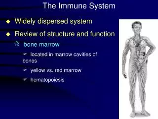
MAST CELLS (MASTOCYTES) • Originate in bone marrow from CD34 progenitor • Basophils may have same progenitor • Development of immature cells at tissue sites • Types • Mucosal • Tryptase production • Development T cell dependent • Connective tissue • Chymotryptase production • Express high levels of IgE receptor
MECHANISM OF TYPE I HYPERSENSITIVITY REACTIONS • FceRI receptor expressed constitutively • Mast cells and Basophils • Activated eosinophils • Allergen binding results in cross-linking of receptors • Cross-linked receptors signal degranulation of cytoplasmic granules • Degranulation results in release and synthesis • Inflammatory mediators, toxins, enzymes
HISTAMINE (BIOGENIC AMINE) • Exerts a variety of physiological effects following binding to specific receptors (H1, H2, H3) • Allergic reactions • Histamine binds to H1 receptors • Physiological effects • Constriction of bronchial / intestinal smooth muscle • Increased permeability of blood vessels • Increased secretion of mucus by goblet cells • Leukocyte chemotaxis
LEUKOTRIENES • Classified as lipid mediators of inflammation • Derived from arachidonic acid via lipoxygenase pathway • Produced by mast cells, monocytes and granulocytes • Leukotrienes (LTA4 – LTE4) • Sustain inflammatory response in allergic disease • Autocrine and paracrine mechanisms • C, D and E are cysteinyl leukotrienes • Increased levels induce anaphylaxis • Physiological effects similar to histamine • More potent / longer lasting than histamine • Vasodilation, bronchoconstriction, neutrophil chemotaxis
PROSTAGLANDINS • Classified as lipid mediators with a variety of physiologic effects • Normal • Inflammation • Derived from arachidonic acid • Cyclooxygenase pathway • Act locally and rapidly metabolized • Produced by all nucleated cells except lymphocytes
CYCLOOXYGENASE PATHWAY • Prostaglandins produced by two different enzymes • Cyclooxygenase-1 (Cox-1) • Cyclooxygenase-2 (Cox-2) • Cox-1 involved in normal physiological functions • Stomach mucus production • Kidney water excretion • Platelet function • Cox-2 involved in inflammatory response
NONSTEROIDAL ANTI-INFLAMMATORY DRUGS (NSAIDS) • Reduce pain, inflammation and fever by inhibition of cyclooxygenase pathway • Non-Selective (Cox-1 and Cox-2 inhibitors) • Acetylsalicyclic acid (Aspirin) • Ibuprofen (Motrin, Advil) • Indomethacin (Indocin) • Naproxen (Naprosyn, Aleve) • Selective (Cox-2 inhibitors) • Celecoxib (Celebrex) • Rofecoxib (Vioxx) • Valdecoxib (Bextra)
EOSINOPHILS • Granulocytic leukocytes (1 – 6% in blood) • Level variation (down in am, up in pm) • Granules • Orange to reddish-orange in color • Uniform in size and evenly distributed • Toxins, enzymes, cytokines and inflammatory mediators • Mature cells reside in • Blood and lower GI tract
EOSINOPHILS • Eosinophil response • Parasites (Helminths) • Main effector cell in allergy and asthma • Induced by drugs, diseases and radiation • Eosinophilia potentially toxic to host • Control mechanism for host toxicity • Limiting bone marrow production • Regulated expression of Fc-epsilon-RI • IgE receptor not expressed in resting state
CASE STUDY – 58 YEAR OLD FEMALE • Presents to family physician with 1 month history • Fever • Cough • Weight loss • Dyspnea • Past and present medical history • Non-smoker • Childhood asthma • Rheumatoid arthritis
CASE STUDY – 58 YEAR OLD FEMALE • Laboratory • CBC with differential • 12,000 leukocytes with 10% eosinophils • Sputum for eosinophils • Unable to produce • Radiology • CXR and CT • Endoscopy • Fiberoptic bronchoscopy with bronchoalveolar lavage (BAL)
CASE STUDY – EOSINOPHILIC PNEUMONIA • Pulmonary eosinophilia or eosinophilic lung disease • Classification • Primary (idiopathic) • Secondary • Parasitic or fungal infection • Immunological or systemic disease • Asthma, HIV, malignancy • Drugs • Antibiotics, NSAIDS, L-tryptophan • Mechanism of disease is unknown
CASE STUDY – 23 YEAR OLD MALE • Presents to family physician with 2 year history of • Dysphagia • Episodes of food impaction • Breads and meats • Past and present medical history • Seasonal allergic rhinitis • Non-smoker
CASE STUDY – 23 YEAR OLD MALE • Laboratory • CBC with differential • 12,000 leukocytes with 11% eosinophils • Endoscopy with esophageal biopsy • Endoscopy showed “ringed esophagus” • Surgical Pathology Report • > 20 eosinophils/HPF (proximal and distal) • Areas of basal cell hyperplasia suggests reflux • No viral CPE, dysplasia or malignancy
CASE STUDY – EOSINOPHILIC ESOPHAGITIS • Etiology is unknown • Associated with food allergy • Milk, eggs, soy, corn, wheat, beef, chicken • Acid reflux • Medications stuck in esophagus • Mechanism • Decrease stretching of esophagus • Treatment is evolving • Prednisone, antihistamines, Mast cell stablizers • Avoidance of implicated food • Proton pump inhibitors (Nexium) ?
BASOPHILS • Granulocytic leukocytes (0 – 1% in blood) • Granules • Violet-blue in color • Variable in size and unevenly distributed • Contents similar to mast cells • Mature cells reside in blood • Basophils similar to mast cells • Constitutive expression of IgE receptor • Significant source of IL-4 • Both interact with eosinophils
IgE MEDIATED ALLERGIC REACTIONS • IgE production is favored following • Chronic exposure to proteins or chemicals bound to proteins • Low molecular weight, soluble, glycosylated proteins • Allergens promote CD4 TH2 response when interleukin-4 is present • Interleukin-4 promotes IgE isotype switch in cognate interaction with B cells • IgE response amplified following release of IL-4 by activated mast cells and basophils
SENSITIZATION TO AN INHALED ALLERGEN • Majority of allergens are components of dried particles derived from plant and animals • Majority of allergens in US are proteases • Cysteine protease in feces from house dust mite • Dermatophagoides pteronyssimus • Papain from papaya fruit • Significance of enzymatic activity of allergens is unknown
GENETIC PREDISPOSITION TO TYPE I HYPERSENSITIVITY • Atopy • Genetic propensity to produce IgE antibodies in response to allergens • Atopic response characterized by elevated levels • IgE and eosinophils • Multiple genes are involved • Chromosome 2 • Regulation of T cell activation • Chromsome 5 • Gene cluster for IL-3, IL-4 and IL-13 • Chromosome 11 • Beta chain of FceRI receptor
TWO STAGES OF TYPE I HYPERSENSITIVITY REACTIONS • Immediate reaction (Stage 1) • Appears within 30 minutes • Subsides within 30 minutes • Late phase reaction (Stage 2) • Appears 6 to 8 hours after immediate reaction has subsided • Subsides within 24 hours • Examples • Wheal and flare (skin) • Breathing capacity (lungs) • Forced expiratory volume in 1 second (FEV1)