OOGENESIS
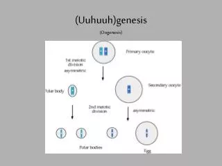
OOGENESIS. Polar bodies. Primary oocyte. Primary spermatocyte. Meiosis I. Meiosis I. Secondary spermatocytes. Secondary oocyte. Meiosis II. Spermatids. Meiosis II. Spermiogenesis. Ovum. Spermatozoa. 1. Four gametes from each primary spermatocyte. 2. Four small gametes of equal size.

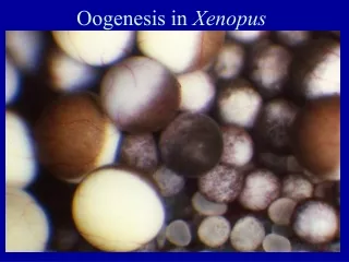
OOGENESIS
E N D
Presentation Transcript
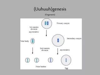
Polar bodies Primary oocyte Primary spermatocyte Meiosis I Meiosis I Secondary spermatocytes Secondary oocyte Meiosis II Spermatids Meiosis II Spermiogenesis Ovum Spermatozoa
1. Four gametes from each primary spermatocyte 2. Four small gametes of equal size 3. Most cytoplasm is shed from spermatocyte 4. Diplotene relatively short 5. Functions in fertilization only after meiosis is complete 3. Cytoplasm conserved in one large gamete - may increase. Thus, in the final gamete there is a large amount of cytoplasm. 1. One gamete from each primary oocyte 2. One large gamete + 2-3 polar bodies 4. Dipotene very long - dictyate state 5. Often functions in fertilization before meiosis is complete Differences between spermatogenesis and oogenesis. Spermatogenesis Oogenesis
Specializations of sperm and egg Spermatozoan 1. Transfer of genetic information to next generation 2. Locomotion 3. Penetration of barriers surrounding the egg 4. Fusion with oolemma 5. Receptor mediated recognition of egg Ovum 1. Transfer of genetic information to next generation 2. Chemoattraction of spermatozoan 3. Prevention of polyspermy 4. Storage of nutrients (importance varies) 5. Storage of cytoplasmic information (importance varies)
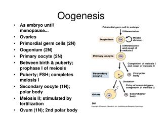
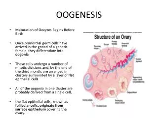
The Dictyate State 1. A sort of “stasis” where the developing oocyte arrests in diplotene of the first meiotic prophase following initial maturation while the mother is an embryo in her mother’s womb. 2. Mediated by a meiosis stabilizing factor that is secreted by the follicle cells of the primordial follicle. 3. At the beginning of a menstrual cycle a number of oocytes in primordial follicles are stimulated by pituitary gonadotropins to continue their maturation. a. Mainly due to leuteinizing hormone (LH) concentration b. Either blocks or deactivates the meiosis stabilizing factor c. As a result, egg maturation continues and meiosis I occurs 4. In many species (including humans), the oocyte then arrests at metaphase of meiosis II until after fertilization.
* All oocytes remain in the
Number of eggs stimulated to begin maturation in a females lifetime. Number of years between puberty and menopause ~ 40 Number of menstrual cycles per year ~ 12 Number of eggs stimulated to continue maturation at each menstrual cycle ~ 9 So, if a woman is never pregnant, the number of eggs she will loose due to the menstrual cycle is about: 40 x 12 x 9 = ~ 4320 eggs Thus, the vast majority of eggs (~495,680) lost during her life degenerate without every being stimulated to continue their maturation.
Follicular development Pictures of follicle stages in digital lab manual.
Storage of energy, raw materials and information in the egg 1. Previtelligenic phase Occurs through early diplotene (during the dictyate state, diplotene may be as long as 50+ years) Lampbrush chromosomes form - RNA transcription including cytoplasmic information for development 2. Vitellogenic phase - starts after the egg is stimulated to continue its maturation Occurs during middle to late diplotene
1. Pre-vitellogenic phase Nucleus swells to form germinal vesicle Pictures of frog follicles in digital lab manual. The nucleus will continue as a germinal vesicle into the vitellogenic phase.
Pre-vitellogenic and vitellogenic phases Frog Lampbrush chromosomes present - RNA synthesis Pictures of lampbrush chromosomes and frog follicle. Chicken http://www.luc.edu/depts/biology/dev/lampbr.htm http://rat.inst.bio.spbu.ru/posters/Paris2001/gsa_p1f1.jpg
Vitelligenic phase - Yolk deposition 1. Vitellogenins - estrogen stimulates synthesis of vitellogenins in liver or equivalent organ. Transported to ovary by circulatory system. Follicle cells may mediate transfer into egg. 2. Molecular structure of vitellogenins modified in the egg. Deposition of yolk in cytoplasm mediated by enzymes, endoplasmic reticulum, golgi bodies, mitochondria. Yolk platelets formed. 3. Vitellins - definitive yolk. Composition - lipid, protein, carbohydrate, phosphorus http://www.luc.edu/depts/biology/dev/vitellog.htm
2. Vitellogenic phase - yolk deposition a. In species with yolky eggs, size of oocyte increases dramatically Pictures of frog follicle stages in digital lab manual.
Egg classification by amount of yolk: 1. Polylecithal, megalecithal - a huge amount of yolk (birds, reptiles, bony fish) 2. Mesolecithal - medium amount of yolk (amphibians) 3. Microlecithal, oligolecithal - very little yolk (most mammals) Egg classification by distribution of yolk: 1. Telolecithal - yolk distributed in gradient, concentrated toward one pole of egg, usually the vegetal pole (e.g. amphibians). 2. Isolecithal - yolk evenly distributed throughout egg cytoplasm (e.g. sea urchins, human) http://en.wikipedia.org/wiki/Image:Salmoneggskils.jpg http://www.exploratorium.edu/cooking/eggs/eggcomposition.html
Cytoplasmic Information Where does it come from? Some of the RNA transcribed from DNA during diplotene of the first meiotic prophase and stored in cytoplasm in inactive form until needed during development. What’s it for? 1. Fast start for development. 2. Can determine fate of specific groups of cells, e.g. primordial germ cells in amphibians and insects.
Ovulation http://www.emc.maricopa.edu/faculty/farabee/BIOBK/femalerepro_3.gif
Ovulation In mammals, the egg is ovulated as a secondary oocyte that is at metaphase of the second meiotic division and is surrounded by layers of cumulus follicle cells. Bovine secondary oocyte surrounded by cumulus follicle cells Secondary oocyte of hampster from which the cumulus cells have been removed by treatment with hyaluronidase. http://www.talbotcentral.ucr.edu/mammalianfert.htm http://arbl.cvmbs.colostate.edu/hbooks/pathphys/reprod/fert/gxport.html
Ovulation [research performed on hampster follicles (Martin et al., 1981; Schroeder and Talbot, 1982)] 1. Enzymes weaken the follicle wall 2. Smooth muscle cells at base of follicle contract 3. This forces the cumulus oophorus containing the oocyte toward the weakened follicle wall, which ruptures 4. The oocyte + surrounding cumulus cells are forced out of the follicle. Arrows indicate weakened follicle wall Arrowheads indicate base of follicle where cumulus + oocyte are or were located http://www.talbotcentral.ucr.edu/mammalianfert.htm http://www.obgyn.net/medical.asp?page=/english/pubs/features/mcgill-student-projects/ovulation-image
Corpus luteum stratum granulosum + theca interna give rise to the corpus luteum. http://www.emc.maricopa.edu/faculty/farabee/BIOBK/femalerepro_3.gif
Picture of cat corpora lutea in digital lab manual. stratum granulosum + theca interna give rise to the corpus luteum.