OOGENESIS
OOGENESIS. By Dr Samina Anjum. Female Reproductive System. PRIMORDIAL GERM CELLS. Gametes are derived from PGCs. Formed in the epiblast during 2 nd week and then move to the wall of yolk sac. Migration of primordial germ cells (PGCs).


OOGENESIS
E N D
Presentation Transcript
OOGENESIS By Dr Samina Anjum
PRIMORDIAL GERM CELLS • Gametes are derived from PGCs. • Formed in the epiblast during 2nd week and then move to the wall of yolk sac.
Migration of primordial germ cells(PGCs) Begin to migrate from the yolk sac in the 4th week and arrive in the gonads by end of 5th week.
In preparation for fertilization germ cells undergo: • Gametogenesis • Oogenesis • Spermatogenesis • Cytodifferentiation
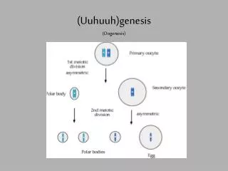
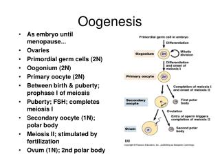
OOGENESIS • Is the sequence of events by which germ cells oogonia differentiate into mature oocytes.
Maturation of oocytes begins: • Before birth. • Continues at puberty. • Ends at menopause.
Morphological changes during maturation of gametes before birth Once PGCs arrive in gonads
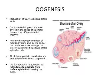
Cont… • After repeated mitotic divisions by the end of 3rd month oogonia arrange themselves in clusters • Surrounded by follicular cells • Majority of oogonia continue to divide by mitosis but some oogonia give rise to primary oocytes that enter the prophase of meiosis I.
Segment of ovary showing different stages of development • By 5th month number of germ cells reaches 7 million in the ovaries. • Cell death (atresia) begins • By 7th month majority of oogonia and primary oocyte degenerate.
At Birth: • No Oogonia present in the ovaries • All surviving primary oocytes have entered the prophase of meiosis I, and most are individually surrounded by flat epithelial cellsto form primordial follicle.
Maturation of oocytes continue at puberty • At birth: The total number of primary oocytes at birth is 600,000 – 800,000 All Primary oocytes are arrested in the Diplotene stage (resting stage during prophase, characterized by lacy network of chromatin) and will not resume 1st meiotic division till puberty is reached. This arrested stage is due to OMI secreted by follicular cells. • At puberty: Number drops to about 40,000 by the beginning of puberty. Rising FSH triggers start of ovarian cycle • Ovarian cycle: Fewer than 500 ovulate in the reproductive life of a female
At puberty a pool of growing follicles is maintained from primordial folliclesEach month 15-20 follicles begin to mature and pass through 3 stages:
Primary Follicle (Preantral stage) • Follicular cells will form a stratified epithelium/granulosa cells around the primary oocyte • Granulosa cells rest on a basement membrane that separates them from ovarian connective tissue (stromal cells) that form theca folliculi. • Zona pellucida- a layer of glycoprotein secreted by granulosa cells and oocyte
Cont… • As the follicles continue to grow, cells of theca folliculi organize into layers. • Finger like processes of follicular cells interdigitates with microvilli of plasma membrane of oocyte
Secondary/Antral/Vesicular FollicleAt maturity size reaches(25mm) • Longest stage • Stratum granulosum 6-12 cell layers • Formation of Antrum: Liquor folliculi (hyaluronic acid) • Granulosa cells surrounding the oocyte remains intact and oocyte is off center. • Well defined Theca interna & externa
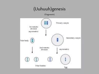
Tertiary or Graafian Follicle(Preovulatory stage lasts for 37 hrs) • Spans entire width of cortex & produces a bulge on the surface of ovary • Large enough to distort the shape of the ovarian capsule • St. granulosum appears to be thinner • One large antral cavity • Cumulus oophorus & corona radiata (loose connection) • A surge in LH, First meiotic division being completed: Primary oocyte divides into a Secondary oocyte and a polar body
Cumulus oophorus • Is a column/mound of granulosa cells that attaches the oocyte to the follicle wall as well surrounds the oocyte. At ovulation, this column of cells is broken or separates to release the oocyte from its follicle attachment. Corona radiata • Is composed of cumulus/granulosa cells that immediately surround the oocyte & send microvilli through ZP that communicate with microvilli of oocyte
mitosis Lies in perivitelline space
Maternal Contributions to the Oocyte As the oocyte is a product of female gametogenesis, the maternal contribution to the oocyte and consequently the newly fertilized egg is enormous. There are many types of molecules that are maternally supplied to the oocyte which will direct various activities within the growing zygote. • Half of zygotic genome • Maternal Mitochondria • Maternal Nucleolus • Maternal Ribosomes
Paternal Contributions to the Oocyte • Half of zygotic genome • Centriole