Cortical Rotation Process and Organizer Formation in Xenopus Oogenesis
170 likes | 217 Vues
Explore the intricate process of cortical rotation in Xenopus oogenesis, where mRNA deposition and cellular movements contribute to organizer formation during early development. Witness the dynamics of Wnt and beta-catenin proteins and their roles in guiding cellular fate through precise cortical movements.

Cortical Rotation Process and Organizer Formation in Xenopus Oogenesis
E N D
Presentation Transcript
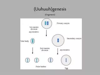
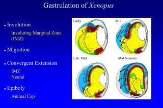
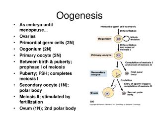
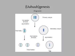
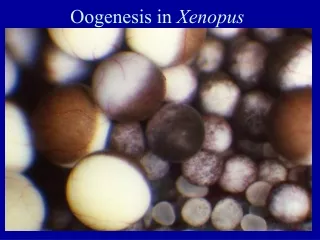
Oogenesis in Xenopus 0.01 mm 1.2 mm Early oocyte, 0.05mm, 1500 nucleoli in nucleus; mitochondrial cloud at vegetal pole Xcat 2 mRNA deposited at vegetal pole cortex VegT mRNA deposited in vegetal hemisphere cortex
Xenopus egg stained with nile blue to see cortical rotation Dye solution is under the grid Done by 20 min after fertilization
Cortical rotation in the Xenopus egg Timed series, surface immobilized, vegetal pole view 30 min 90 min Double stained egg: surface is yellow, interior is green 30° movement
During cortical rotation, a thin layer of aligned microtubules forms between the cortex and core: the tracks for kinesin motors moving the cortex and vesicles. Vegetal pole, 10 um into the egg, 60 min after fertilization.
Cortical rotation: Movement of wnt11 and vg1 mRNAs and proteins from the vegetal pole to one side (where the organizer will later form) Grey crescent wnt11 mRNA and protein (b-catenin stabilizing agent) wnt11 mRNA and protein (b-catenin stabilizing agent)
Planar microtubule array, plus ends aligned, on surface of cytoplasmic core DORSAL-grey crescent side VENTRAL-sperm entry side
Rotation of cortex over core, by 30 degrees, revealing grey crescent Sperm entry side Grey crescent • Before rotation After rotation
Wnt11 protein Outside cell Inside cell Axin b-catenin builds up b-catenin Wnt is present. b-catenin protein is not degraded. It builds up, binds to Tcf/LEF and enters the nucleus. Wnt is absent. b-catenin mRNA is continuously translated and the protein is continuously degraded.
During cortical rotation, beta-catenin protein accumulates on the dorsal side of the egg (fluorescent antibody staining)
Beta-catenin protein is stabilized on the grey crescent side until the late blastula stage, when the organizer is formed. 4000 cell blastula shown. As nuclei form, beta- catenin protein enters D D V D D Beta-catenin protein detected by a specific antibody.
Change cortical rotation, change development Normal rotation, normal organizer (60° wide), normal development Partially dorsalized Prevent microtubule polymerization, get no rotation Too many microtubules, or LiCl Dorsalized embryo Excess organizer Ventralized embryos-no organizer
Artificial rotation, forced by gravity, rescues eggs that failed to do their own rotation.
Two artificial rotations, forced by gravity, give two organizers, and twins.