CHAPTER 3 Cell Structure and Function
CHAPTER 3 Cell Structure and Function. Life has a cellular basis. All organisms, including humans, are composed of cells. There is no smaller unit of life that is able to: reproduce, respond, grow & develop, and take in and use materials from the environment .

CHAPTER 3 Cell Structure and Function
E N D
Presentation Transcript
Life has a cellular basis. All organisms, including humans, are composed of cells. There is no smaller unit of life that is able to: reproduce, respond, grow & develop, and take in and use materials from the environment. The work of 17th century scientists who had invented early microscopes and those that used them led to one of the fundamental principles of modern biology, the Cell Theory. Cells
Cells are the basic working unit of life. 2. All living things are made up of one or more cells. 3. New cells ariseonlyfrom pre-existing cells. The Cell Theory States:
Cell Size The ratio of surface-area-to-volume explains the small size of cells. Nutrients enter a cell and wastesexita cell at its surface. The ability to get more materials in and out depends on a greater amount of surface area. Volume represents cell needs.
Cell Size Proportionally as a cell grows it becomes larger in volume andsurface areadecreases. There is a limitto how large an actively metabolizing cell can become. Cell division restores the amount of surface area neededfor adequate exchange of materials.
Microscopes Light microscopes • Use glass lenses & light rays to obtain images • Better suited for living specimens • Lower magnification Electron microscopes • Use electrons to obtain images • Obtain higher magnification and resolution Microscope use and its parts will be studied in lab.
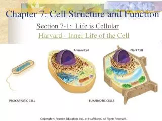
How Cells are Organized Cells can be classified as either prokaryotic or eukaryotic. Prokaryotic cells contain no membrane-bound organelles and lack anucleus. They have a plasma membrane and contain cytoplasm; have ribosomes, and a ring of DNA. Most prokaryotes are bacteria.
How Cells are Organized You and nearly all other life forms that you experience with your unaided eyes are eukaryotes. The vast majority of eukaryotes that we knowingly interact with each day, mainly land plants and animals are large organisms, usually consisting of trillions of cells.
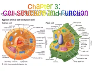
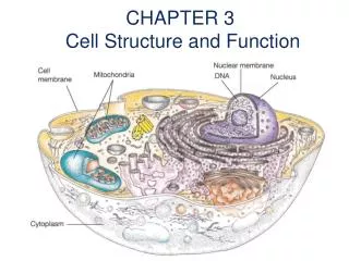
How Cells are Organized Almost all eukaryotic cells have 3 main areas • An outer membrane called the plasma membrane. • A semifluid medium inside called thecytoplasm. The substance contains various organelles(small specialized structures). 3. A large central membrane-enclosed nucleus, where DNA is found.
Cellular Organization Nucleus Central sphere of the cell Plasma Membrane Outer boundary between the cell and the environment Cytoplasm Everything between the plasma membrane and the nucleus Organelles Specialized structures in the cytoplasm
The plasma membrane is the boundary between the outside and inside of the cell. The plasma membrane is a phospholipid bilayer with attached or embeddedproteins. At body temperature the bilayer is a liquid. This allows the proteins to move about. This structure of being liquid and having shifting proteins is described as the fluid-mosaic model. Plasma Membrane - Structure
Short chains of sugars attach to the proteins and lipids and are called glycoproteins and glycolipids. They mark cells as belonging to a particular individual. A common example of this can be seen in the different blood types of people. Plasma Membrane - Structure
Plasma Membrane - Functions The plasma membrane keeps the cell intact. The plasma membrane allows only certain molecules to enter and exit freely. Therefore, the plasma membrane is said to be selectively permeable.
Passive Transport Passive transport is a way for molecules to enter or exit a cellwithoutusingcellular energy. Diffusionis the random movement of molecules from an area of higher concentration to an area of lower concentration until they areequally distributed.
Osmosis Osmosis is the diffusion ofwateracross a semi-permeable membrane.
Tonicity Tonicity is the term used to describe the solute concentration in a solution surrounding a cell. Example: 2% salt solution (98% water)
Solution Types - Identified by Tonicity Isotonic – Solution outsideof the cell is equal in concentration of water and solute. This causes cells to: Neither gain nor lose water.Normal cell size and shape maintained. Hypotonic – Solution outside of the cell with a higher concentration of water and lower solute. This causes cells to:Gain water. Cells increase in size or burst.
Solution Types - Identified by Tonicity Hypertonic – Solution outside of the cell has a lower concentration of water and higher solute. This causes cells to:Lose water. Cells shrink or shrivel in size. Osmotic pressure controls water movement in our bodies. Examples: Absorption in the small and large intestine and in the kidneys.
Facilitated Transport The transport of solutes from high to low concentration by means of protein carriers within the membrane. This transport is also without using cell energy.
Active Transport Occurs when a molecule is moving from a lower to higher concentration. Movement requires a protein carrierand the use of cellular energy. Protein carriers act like a pump to move molecules against their concentration gradient (low to high).
Endocytosis Endocytosisoccurs when a portion of the plasma membrane forms a pouch to surround asubstanceandfluid. The membrane pinches off and forms avesicle.
Types of Endocytosis When cells like white blood cells do endocytosis and the membrane pushes out to engulf a particle like a bacterium, the process is calledPhagocytosis. Pinocytosisis taking insmall moleculesand fluid. The membrane invaginates forming a vesicle.
Exocytosis The process is calledExocytosiswhen a vesicle fuses with the plasma membrane to move substancesout of a cell.
The nucleus and several organelles are involved in the production and processing of proteins. The nucleus is the largest structure in the cell, usually spherical, found near the center and stores genetic information. Segments of DNA threads called genes contain information for the production of specific proteins. The nucleus is thus called thecontrol center of the cell.
Threads found inside are made of DNA and protein and are called chromatin. These threads are immersed in a semifluid medium called nucleoplasm. Thenucleolus is a dark dense region where ribosomal RNAis produced and joins with protein to form subunits calledribosomes.
Thenuclear envelopeseparates the nucleus from the cytoplasm and is perforated with passageways callednuclear pores. Nuclear pores Nucleoplasm Nucleous Nuclear Envelope Endoplasmic reticulum
Ribosomes Ribosomes are tiny granules composedproteinsandrRNA. Ribosomes are whereprotein synthesisoccurs. Ribosomes are often attached to membranes in the cytoplasm called theendoplasmic reticulum (ER). Ribosomes can also be found alone in the cytoplasm or in groups calledpolyribosomes.
The Endomembrane System The endomembrane system consists of the: Nuclear envelope Endoplasmic reticulum Golgi apparatus Lysosomes Vesicles
The Endoplasmic Reticulum The endoplasmic reticulum (ER) is a system of membranes within the cytoplasm and continuous with the nuclear envelope. Two types are: Rough ER is studded with ribosomes on the cytoplasm side. Proteinsare synthesized here.
The Endoplasmic Reticulum Smooth ERlacksribosomes and is the site of synthesis of phospholipids and other substances. The ER forms vesicles, which are membrane sacs used to transport substances.
Endoplasmic Reticulum Smooth ER Rough ER Ribosomes
The Golgi Apparatus The Golgi apparatus consists of a stack of curved saccules. The Golgi apparatus receives protein and lipid-filled vesicles from the ER. Ends of the saccules expand forming vesicles, which leave the Golgi apparatus. The Golgi apparatus processes, packages, secretes, and distributessubstances within and out of the cell.
Lysosomes Lysosomes are vesicles produced by the Golgi apparatus. Lysosomes contain hydrolytic enzymes. All body cells have lysosomes, but certain white blood cells have many and use them to help protect us from disease-causing microbes.
Lysosomes Lysosomes can fuse with vesicles to digesttheir contents. They engulf, digest, and release simpler substances into the cytoplasm for reuse.
The Cytoskeleton The cytoskeleton is a network of protein fibers that crisscross within the cytoplasm. The cytoskeleton is made up of different types of fibers that help maintain a cell’s shape and can anchor the organelles or assist with their movement.
Cell Movement Cilia, tiny hairlike structures on cell surfaces and Flagella, large single projections from cells are involved inmovement.
Cilia and Flagella Cilia that line the respiratory tract sweep debris trapped in mucus out and cilia in the oviducts move eggs along. Spermcells have flagella that move them to find and fertilize eggs.
Mitochondria Structure Mitochondriaare found in plant and animal cells. Mitochondria are bounded by a double membrane. The inner membrane is folded to form shelves called cristae. The folds project into the matrix an inner space filled with a gel-like fluid that contains enzymes, which are used to break down glucose products.
Additional Mitochondrion Facts Mitochondria were originally prokaryotes. Mitochondria contain their own DNAand ribosomes. Mitochondria are only produced by other mitochondria. In humans and most multicellular organisms mitochondria are maternal inherited from the mothersegg.
Mitochondria Function Mitochondria are often called the powerhouses of the cell. Mitochondria convert the chemical energy ofglucoseinto the chemical energy ofATP. This cell process is called cellular respiration.