Download

1 / 89
1k likes | 1.49k Vues
Lecture 4 and 5 Leukemia and Chronic myeloproliferative disorders Abdulkarim Aldosari. O bjectives. Define leukemia Compare and contrast acute versus chronic leukemia, and leukemia versus lymphoma Compare and contrast acute myeloid and acute lymphoblastic leukemia

E N D
Lecture 4 and 5 Leukemia and Chronic myeloproliferative disorders AbdulkarimAldosari
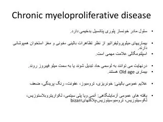
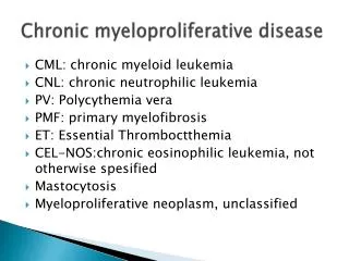
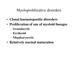
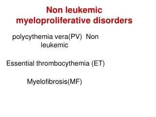
Objectives • Define leukemia • Compare and contrast acute versus chronic leukemia, and leukemia versus lymphoma • Compare and contrast acute myeloid and acute lymphoblastic leukemia • Describe FAB and WHO classifications for ALL and AML • Describe characteristic morphology and cytochemical staining patterns for each of the subtypes of AML • Correctly identify blast on a peripheral smear and distinguish between the features of a lymphoblast and a myeloblast • Name and describe the characteristics of each of the chronic myeloproliferative disorders
Definitions and Overview • Neoplasms = “new growth” due to dysregulated proliferation of a single transformed cell • External growth factors that regulate proliferation are reduced or eliminated due to genetic mutations in the transformed cell • Benign neoplasm = differentiated cells that do not spread or invade surrounding tissue • Can progress with further mutations to a malignant neoplasms • Malignant neoplasms = “deadly” “having the potential to produce death” • Proliferating cells with potential to metastasize • Only malignant tumors are correctly termed as cancer • Cancer is actually a malignancy of epithelial tissue, it is also commonly used to include all malignant neoplasms.
Definitions and Overview • Leukemia = lymphoid and myeloid malignant bone marrow neoplasms • when abnormal cells are seen in both the bone marrow and the peripheral blood • Lymphoma = abnormal proliferation of lymphoid cells within the lymphatic tissue or lymph nodes • Spectrum
Leukemia – definition and overview • A malignant disease that affects the hematopoietic tissue • Normal bone marrow is replaced by abnormal blood cells (neoplastic cells) • Cells sometimes found in the PB, in the reticuloendothelial organs, and other organs • Prognosis – poor > death • Incidence of leukemia in the USA = 8-10 new cases/100,000 individuals per year • Approximately every 10 minutes, someone in the US dies from a blood cancer= nearly 150 people each day or more than six people every hour.
Leukemia statistics • There are an estimated 310,046 people living with, or in remission from, leukemia in the US. • In 2013, 48,610 people are expected to be diagnosed with leukemia. • In 2013, 23,720 people are expected to die from leukemia. • Approximately 33 percent more males are living with leukemia than females. • More males than females are diagnosed with leukemia and die of leukemia. • Leukemia causes almost one-third of all cancer deaths in children and adolescents younger than 15 years. • www.lls.org/diseaseinformation/getinformationsupport/factsstatistics
Leukemia – definition and overview • Acute leukemia = rapid onset with abnormal expansion of immature cells/blasts • Chronic leukemia = slow progression with abnormal expansion of mature cells • Divided by two cell types: myeloid and lymphoid • Acute myeloid leukemia (AML), or chronic myeloid leukemia (CML) • Acute lymphoid leukemia (ALL), or chronic lymphoid leukemia (CLL) • Chronic leukemia (CML or CLL) generally associated with adults • Most common form in children = ALL
Leukemia – definition and overview • Most cases affect adults = 10:1 compared to children • Most common = AML (34% of cases) and CLL (29% • CLL is extremely rare in children – unusually before the age of 40 • 50% of all cases occur after the age of 64 • Adults 70-90 yrs. old > CML • Higher rates in males than females • Higher in European descent than African, lowest in American Indians and Alaskan
Comparison of acute and chronic leukemia Acute – Affects all ages, sudden onset Becomes fatal in 6 months if untreated Loss of BM function Mild to severe anemiaand/or thrombocytopenia, WBC count- variable Immature neoplastic cells Chronic - Affects mostly adults, can last 2-6 years, Early diagnosis = ↑ survival Elevated WBC count > 50,000/µL Mature neoplastic cells Mild anemia, normal platelet count, Prominent and massive organomegaly
Advances • Advances in diagnosis and treatment > improvement in survival • Laboratory analysis – cytochemical cytogenetic, immunologic, molecular techniques > id specific categories of leukemia > distinct treatment protocols • Bone marrow and stem cell transplantation • Cytotoxic drugs, radiation • Targeted approaches – tyrosine kinase inhibitors, protease inhibitors • Genetic mutations - altered expressions of oncogenes and tumor suppressor genes > unregulated cellular proliferation
Factors that predispose or increase the incidence of leukemia Host Factors Hereditary – congenital chromosomal disorders Abnormal chromosomal number Immunodeficiency Chronic marrow dysfunction Environmental Factors Radiation Chemicals Drugs Viruses
Classification of leukemias Classified according to cell type - cell maturity and/ or cell lineage French-American- British (FAB) World Health Organization (WHO) • Divide leukemias into ALL and AML
Acute leukemia ALL more common in children = 75% of childhood leukemias AML more common in adults = 80% Incidents of AML increases with ageMedian age 63 yrs.
Clinical onset of acute leukemia • Within a few weeks pts. present with weakness, bleeding abnormalities, flu-like symptoms • Due to proliferation and accumulation of abnormal cells > • BM failure > anemia, Granulocytopenia, thrombocytopenia and their sequelae • Organ infiltration > marrow expansion, spleen, liver, lymph nodes, CNS, gums, mouth Symptoms • Bleeding, DIC, infections, gingival hypertrophy = swelling of the gums, oral lesions • Bone and joint pain, neurologic conditions - CNS > intracranial pressure> nausea, vomiting, headache
Laboratory evaluation of acute leukemia Clinical history, physical examination, CBC and PB smear • Mild anemia – normocytic normochromic • ↓ platelet count • Variable WBC • Blasts or other immature cells (may be rare or absent) • nRBC • Myelodysplastic features - Pseudo-Pelger-Huet • Hypogranular neutrophils Diagnosis can be established with PB smear but BM is the preferred specimen
Laboratory evaluation of acute leukemia BM aspirate and biopsy – required to establish diagnosis • >20% blast in BM or PB = minimum WHO classification requirement for acute leukemia • >30% for FAB classification Morphological examination – cell lineage, guide to further studies Immunologic cell markers studies – flow cytometry for blast immunophenotype Cytochemical staining – differentiate granulocytic from monocytic leukemias Karyotyping Molecular studies DNA flow cytometry Electron microscopy
Specimen and evaluation of morphology Properly collected samples - In EDTA tubes Adequate amount of BM material collected and prepared Morphology • Where cells are not overcrowded, or shape distorted • Distinguishing features of myeloid and lymphoid cells • Size, Nuclear chromatin, cytoplasm, nucleoli • Auer Rods – (+) in 60% of AML patients
Specimen and evaluation of morphology Cytochemical staining • Identify chemical components of the cells – enzymes or lipids • Distinguish between AML and ALL • Sub-classify AML • Fresh preparations preferred, control smears • Look for (+) reaction in leukemic blasts rather than in mature cells
Cytochemical reactions Myeloperoxidase (MPO) Stains primary granules • Myeloperoxidase is most abundantly expressed in neutrophil granulocytes • Myeloperoxidase staining to differentiate AML from ALL has been supplanted by the widespread use of flow cytometry • More specific than Sudan Black B • The red reaction = myeloid leukemia • Fresh sample required
Cytochemical reactions Sudan Black B (SBB) Stains primary and secondary granules • Stains phospholipids, neutral fats, sterols • Most sensitive for granulocytic precursors • Less specific than MPO • Does not diminish with time • Useful for samples that are not freshly drawn
Cytochemical reactions Nonspecific esterase (alpha-naphthyl butyrate) (NSE) Most specific for monocytes • Monocytes and their precursors stain a diffusely (+) pretty red color, T-lymphocytes may have some dot-like (+)staining, may also be (+) in megakaryocytes • Neutrophils and their precursors and most of the other types of cells in the bone marrow are negative Specific esterase (chloroacetate esterase CAE) • (+) for myeloblasts
Cytochemical reactions Periodic acid-Schiff (PAS) Stains glycogen, glycoproteins, glycolipids Does not distinguish between ALL and AML • (+) Lymphocytes – granular pattern • (+) Granulocytes – diffuse staining pattern • (-) Normal erythroid precursors • (+) Erythroblasts Differentiate erythroleukemia from pernicious anemia Erythroleukemia - rare form of acute myeloid leukemia where the myeloproliferation is of erythroblastic precursors. PA- Megaloblastic anemias = blood smear shows large, fragile, immature erythrocytes, known as megaloblasts
Cytochemical reactions Acid Phosphatase • Constituent of lysosomes present in most cells • Characteristic for T cell ALL > focal polarized acid phosphatase activity • (+) Hairy cell leukemia uninhibited by tartrate resistant acid phosphatase • Other leukocytes inhibited by tartrate Terminal deoxynucleotidyl transferase (TdT) • Nuclear enzyme that differentiates ALL from AML • DNA polymerase present in both T and B lymphocyte progenitors, • Absent in normal myeloid cells • Performed by flow cytometry, immunofluorescent microscopy, immunohistochemical methods
Immunologic markers Antibodies used to identify specific antigens characteristic to a particular cell line Antigens present on blast cell surface or within the cytoplasm Two techniques: • Flow cytometry – done on PB and BM aspirate • Immunohistochemistry – paraffin sections of core biopsy • Paraffin section - a histologic section cut from tissue that has been embedded in paraffin wax
Cell surface markers • Different cell lines and different stages express different surface proteins • Highly specific monoclonal antibodies - discriminate various stages of human lymphocyte and granulocyte differentiation (see table 16-9) • CD = cluster designation – a protocol used for the identification and investigation of cell surface molecules • provide targets for immunotyping cells • Used when leukemic cells are poorly differentiated by cytochemical stains • Replacing conventional cytochemical methods • Use a panel of markers that includes abs to several myeloid-associated antigens No single marker defines all forms of AML
Cell surface markers • Fresh sample with viable cells required to prevent nonspecific staining • Immunofluorescent staining + analysis with flow cytometer • Staining can be observed with a fluorescent microscope
Cytoplasmic markers Markers directed at cytoplasmic antigens For flow cytometry – additional step to fix cells to allow abs to enter the cytoplasm Quantity of surface ags and cytoplasmic ags varies Especially useful in assessing cell lineage in ALL CD3 – T-cell ALL CD22, CD79a – aid in defining B-cell lineage In ALL Cytoplasmic IgM heavy chain Pre-B-cell ALL (+) Early-pre-B-cell ALL (-) Pre-B-cell ALL has worse prognosis than early-pre-B-cell ALL Cytoplasmic heavy chain staining no longer routinely preformed > FISH now used
Cytogenetics Cytogenetics refers to the microscopic analysis of chromosomes in individual cells • Play a role in diagnosis • Sub-classification of leukemia • Prognosis • Selection and monitoring of therapy Cytogenetics studies can be performed on • fresh blood • bone marrow • prenatal specimens • solid tissue specimens • fixed specimens
Cytogenetic analysis FISH –(fluorescent in situ hybridization) • Fluorescently labeled DNA probes that are capable of hybridizing to complementary chromosomal regions • To view the chromosomal location of a particular gene or DNA sequence through a microscope • Used to identify a translocation of chromosome 1 to 19 - t(1;19) and other cytogenic abnormalities • Translocation t(1;19) in pre-B-cell ALL linked to poor prognosis of pre-B-cell ALL • 2nd most common structural abnormality • Translocation = reciprocal interchange of portions of two chromosomes,
Cytogenetic analysis Karyotypeis a test to identify and evaluate chromosomes • Metaphase chromosomes +stains = distinctive banding patterns→ chromosome pairs are then arranged into a standardized format known as a karyotype. • Hybridization probe corresponded to a segment of chromosome • Permits the simultaneous tracking of all human chromosomes • Detect chromosomal rearrangements; translocations, deletions, duplications, isochromosomes • Fails to detect chromosomal abnormalities in 40-50% of AML • Automated spectral karyotyping has ↑ the sensitivity and accuracy of karyotypic analysis
Cytogenetics Chromosomal abnormalities associated with different forms of leukemia • Philadelphia chromosome t(9;22) > CML • Translocation t(15;17) > acute promyelocytic leukemia (M3) • t(8;21) > M2 – favorable prognosis, good response to treatment • Table 16-10
Molecular genetics Cloning of genes: Ig genes, T-cell receptor genes, other genes DNA FISH probes PCR assays – more sensitive than FISH Microarray assays
FAB classification of AML M0 through M7 • based on the type of cell from which the leukemia developed • how mature the cells are • based on how the leukemia cells looked under the microscope after routine staining M0 through M5 all start in precursors of white blood cells M6 starts in very early forms of red blood cells M7 starts in early forms of cells that make platelets Subtypes are linked with certain symptoms • Bleeding or blood clotting problems are often a problem for patients with the M3= acute promyelocytic leukemia (APL).
Alternate names for AML • acute nonlymphoblastic leukemia (ANLL • acute myelogenous leukemia • acute myeloblastic leukemia • acute granulocytic leukemia
Main features of AML M0 • 5% of adult AML • (-) for all cytochemical stains • poor prognosis compared to other types M1 • 15% • Aggressive leukemia M2 • 25% - most common type
Main features of AML M3 • 10% • Auer rods present • Bleeding or blood clotting problems (DIC) • t(15:17) M4 • 20% • (+) NSE • Myeloid and monocytic antigens M5 • Excess of monocytic cells • Skin and gum involvement
Main features of AML M6 • 5% • Erythroid precursors • Megaloblastic, ineffective erythropoiesis, ringed sideroblast • Difficult to differentiate from Myelodysplastic syndromes • M6 = ≥ 50% erythroblasts and myeloblasts > 30% M7 • 5% • Proliferation of megakaryoblasts
WHO classification Newer system that includes factors that help to better classify cases of AML based on a patient’s outlook AML with recurrent genetic abnormalities • AML with a translocation between chromosomes 8 and 21 • AML with a translocation or inversion in chromosome 16 • APL (M3), which usually has translocation between chromosomes 15 and 17 • AML with changes in chromosome 11 AML with multilineage dysplasia • more than one abnormal myeloid cell type is involved AML related to previous chemotherapy or radiation
WHO classification AML not otherwise specified • Includes cases of AML that don’t fall into one of the above groups • Similar to the FAB classification • Undifferentiated AML (M0) • AML with minimal maturation (M1) • AML with maturation (M2) • Acute myelomonocytic leukemia (M4) • Acute monocytic leukemia (M5) • Acute erythroid leukemia (M6) • Acute megakaryoblastic leukemia (M7) • Acute basophilic leukemia • Acute panmyelosis with myelofibrosis • Myeloid sarcoma • also known as granulocytic sarcoma or chloroma
Treatment of AML and response to treatment Cytogenetic and molecular abnormalities provide best information for predicting clinical outcome and therapy Subgroups to determine therapy • Acute promyelocytic (APL) -DIC > risk of bleeding • AML with inv(16) or t(8;21) • AML in the elderly Targeted drug treatments • Based on genetic profile
Acute lymphoblastic leukemia • 75% of ALL occur in children • Precursor B-cell ALL • Usually fatal > 80% may be cured • Classification based on immunophenotypic, molecular, cytogenetic and morphologic characteristics • WHO - immunophenotypic, molecular, cytogenetic • FAB – blast morphologic characteristics
B-cell development TdT – terminal deoxynucleotidyl transferase = nuclear enzyme
FAB classification of ALL Separated into three morphological groups L1 • Small uniform lymphoblasts • Scanty basophilic cytoplasm • Variable vacuoles • Inconspicuous nucleoli • Regular nuclear shape, occasional clefting • Homogenous chromatin • N:C - high
FAB classification of ALL L2 • Large heterogeneous/pleomorphic blasts • Moderate cytoplasm, often intensely basophilic, • Variable vacuoles • Large nucleus, irregular shape with clefting and indentation, • Large nucleoli • Variable nuclear chromatin • N:C – lower than L1
FAB classification of ALL L3 • Medium to large homogenous cells • Moderate cytoplasm that is intensely basophilic • Prominent cytoplasmic vacuoles • At least one prominent nucleoli (may be 2-4), • Round to oval nucleus • Finely stippled homogenous chromatin • Cytologically identical to Burkitt’s and Burkitt’s like lymphoma • Has mature phenotype (i.e. expresses surface immunoglobulin) • Fat vacuoles are Sudan black+, Oil red O+ and PAS • L3 was considered worst prognosis based on morphology
Burkitt’s Leukemia/Lymphoma (Mature B-Cell ALL) • Similar to L3 but lacks the immunophenotypic characteristics of early B cell • The TdT is negative; CD19, CD20, HLA-DR (+); most cases (-) CD10 • WHO classification does not include the FAB L3 subtype • Burkitt leukemia is rare • Characterized by rearrangement of the c-mycgene on chromosome 8 by one of three chromosomal translocations – t(8;14), t(2;8), t(8;22) • Poor prognosis, worse than pre-B ALL