Download
1 / 28
330 likes | 581 Vues
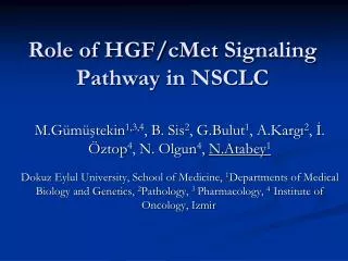
Role of HGF/ cMet Signaling Pathway in NSCLC. M . G ümüştekin 1 ,3,4 , B . S is 2 , G . B ulut 1 , A . K argı 2 , İ . Ö ztop 4 , N . O lgun 4 , N . A tabey 1

E N D
Role of HGF/cMetSignalingPathway in NSCLC M.Gümüştekin1,3,4, B. Sis2, G.Bulut1, A.Kargı2, İ. Öztop4, N. Olgun4, N.Atabey1 Dokuz Eylul University, School of Medicine, 1Departments of Medical Biology and Genetics, 2Pathology, 3 Pharmacology, 4 Institute of Oncology, Izmir
Receptor Tyrosine Kinases • HGF-c-Met signaling pathway • Importance and role in carcinogenesis • Role in development of lung cancer • Probable role in NSCLC ? • IHC data of of c-Met, HGF ve some of target genes • Mutation analysis of Exon 14
Protein Tyrosine Kinases • Enzymes that catalyse transfer of a -phosphoryl group from ATP to serine, treonine or tyrosine residues of other proteins • Normal cell and tissue development • Increased RTK activity • Proliferative diseases *Solid Tumors *Leukemia and Lymphomas *Pseurosiasis, etc
Protein Tyrosine Kinases • 20 subfamilies according to catalytic tyrosine kinase domain homology • Extracellular ligand binding region • Single transmembranal region • Intracellular tyrosine kinase region • Conserved Ig-like and EGF-like regions rich in sistein
Intracellular Region • Extracellular Domain • Upon ligand binding, receptor dimerisation and changes in confirmation, leading to increase in kinase activity and autophosphorylation • Catalytic Domain: Highly conserved • ATP binding region that catalyses receptor autophosphorylation and tyrosine phosphorylation of RTK substrates
Activation of Receptor Tyrosine Kinases Autophosphorylation has dual effect: 1- Activation following Tyr phosphorylation leads to phosphorylation of Tyr away from the active site 2- These phosphorylated residues creates binding regions for the effector molecules (those containing SH2 ve PTB domains)
Effectors of Receptor Tyrosine Kinases • PI3K p85 subunit • Non-receptor PLCgama • Src family tyrosine kinases • p120GAP (a GTPase activating enzyme in Ras signaling • Grb2 Adaptor protein • SH-PTP2 (Shp2) Tyr specific protein phosphotase
Plazma Zarı c-Met Reseptor Tyrosine Kinase S: “Sema” domain C:sistein-rich domain Ig: immunoglobulin domains K: kinase domain HOS, 7q21-q31, 12 kb, 150 kDa
Substrate binding regions of c-Met and Tpr-Met Ekstraselüler Domain Transmembran Domain Juxtamembran Domain Kinaz Domaini Karboksi terminal “docking” bölgesi c-Met Tpr-Met
Mitogen Morphogen Motogen Hepatocytes Epithelial cells Endothelial cells Neurons Melanocytes Lymphocytes Bone marrow derived cells Hepatocyte Growth Factor / Scatter Factor (HGF/SF)
Normal development and adult homeostasis Development og kidney, liver, spleen and placenta Neuronal development Branching morphogenesis Kidney and lung regeneration Normal functions of liver Wound healing Carsinogenesis Carcinomas (kolon, mammary, head-neck, gastric, lung, thyroid and renal carcinomas) Melanomas Sarcomas Lung?? HGF/SF
N: N terminal Domain,K1-K4 Kringle Domains, SPH:Serin Proteinase Homology Domain Hepatocyte Growth Factor / Scatter Factor (HGF/SF)
Nonparanchimal cells Pro-HGF (latent) 92 kDa HGFA (active) Trombin HGF (Aktive) 69kDa+32 kDA HAI HGFA (latent) c-Met (HGF receptor) Target Cells Hepatocyte Growth Factor / Scatter Factor (HGF/SF)
Unstimulated Stimulated Plasma Membrane Substrates Effect of HGF stimulation
*Cell polarity *Actin cytoskeleton *Motility *C/C contacts *Migration *Invasion *Survival *Proliferation *Cell Cycle regulation Branching morphogenesis HGF-c-Met Signaling Pathway Plasma Membrane
HGF/c-MetInvasion Metastsis • *HGF is the unique extracellular signaling molecule that can trigger increase in extracellular matrix proteolysis, cell dissociation, cellular motility • *Inhibitors that block this pathway blocks cell motility, invasion and angiogenesis as well. • Atabey N, et al. J of Biol Chem 276 (17) : 14308-14, 2001, • Maulik G et al, Cytokine Growth Factor Rev 13(1): 41-59, 2002, • Soriano JS, et al Mol Cancer Ther, 2004
Brain Liver Bone Bone Marrow Normal Epithelial Cells Transformed cells Motility Scattering Migration Invasion Metastasis Organ spesific Metastasis ECM VEGF, bFGF HGF, uPA,MMPs Relationship between c-Met-HGF Signaling and Solid Tumor Metastasis
C-Met in NSCLC • Increase in c-Met expression and autonomous Met kinase activity • In patients exhibiting recurrence, HGF level is high, related with poor prognosis Siegfried JM et al, Cancer Res 57(3):433-9, 1997. • Constitutive and paracrine activation of c-Met activates signaling pathways that regulates cell survival and proliferation. This leads to tumor progression. Qiao H et al, J Cell Biochem 86(4): 665-77, 2002. • Adenokarsinomda %35, Büyük hücreli andiferansiye kanserinde %20, Skuamöz hücreli kanserde ise normal akciğer dokusundan daha az veya yakın oranda, Adenokarsinomda c-met’in ekspresyon düzeyi ile tümör diferansiasyonu arasında korelasyon var Tsao MS et al, Lung Cancer 20(1): 1-16,1998.
HGF and c-met expression analysis in 63 NSCLC tissue samples obtained from Dokuz Eylul University, School of Medicine, Department of Pathology was established immunohistochemically.
Due to IHC analysis, of these NSCLC tissue samples • c-Met expression was increased in 81%, • HGF expression was increased in 48%. c-Met HGF
In late stage cases, c-Met expression was high in 72 %, HGF expression was high in 58 % of the cases. • There was no corelation between tumor size, lymphatic metastasis, tumor stage and recurrency. • In 3 cases with metastasis, c-met expression was found to be high.
Method Paraffin blocked tissue samples DNA Isolation (Nucleospin) Polymerase Chain Reaction (PCR) Agarose Gel Electrophoresis DNA sequence analysis (ABI Prism) Blast analysis (Multalin)
with primers flanking exon 14 • c-Met reseptörünün tirozin kinaz bölgesini kodlayan 14.eksona özgü primerler • Forward primer: GCCCATGATAGCCGTCTTTA • Revers primer: CAACAATGTCACAACCCACTG
Results Exon 14 PCR Amplification DNA Sequence Analysis 256 bp Squamous cell Ca Ca T3N0M0 c-met overexpression A deletion Frameshift
1- Mutation analysis of exon 13 and exons 15-20 2- Increase sample group size3- Identification of molecules in HGF/c-Met signaling that may participate in NSCLC development Studies under progress