Download
1 / 13
130 likes | 152 Vues
Learn the principles and procedures of blood glucose testing using a spectrophotometer in the laboratory with this comprehensive guide. Understand specimen collection, storage, and interpretation of glucose levels. Explore micropipettor usage and maintenance tips.

E N D
Glucose test Ms. Ibtisam alaswad Ms. Nour A. taim
Introduction Blood glucose :_ • The main sugar that the body makes from the food in the diet. Glucose is carried through the bloodstream to provide energy to all cells in the body. Cells cannot use glucose without the help of insulin. • Glucose is a simple sugar (a monosaccharide). The body produces it from protein, fat and, in largest part, carbohydrate. Ingested glucose is absorbed directly into the blood from the intestine and results in a rapid increase in blood glucose. Glucose is also known as dextrose.
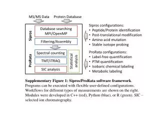
The Objectives to be achieved • To understand the principle to spectrophotometer A spectrophotometer is used to measure the amount of light a sample (be it liquid or solid) absorbs. Light is passed through the sample and a detector measures the intensity of that light that passes through the sample and hits that detector. • To determine the blood glucose concentration as an application of spectrophotometer use • To know how to use some devices in the lab
The principle of Glucose test Glucose present in the plasma is oxidized by the enzyme glucoseoxidase (GOD) togluconicacid with the liberation of hydrogen peroxide, which is converted to water and oxygen by the enzymeperoxidase (POD). 4_aminophenazone, an oxygen acceptor, takes up the oxygen and together with phenol forms a pinkcolouredchromogenwhich can be measured at 515nm.
Specimen type, collection and storage • Plasma is the specimen of choice for glucose estimation. Plasma glucose levels have been checked to be quite stable for 6 hours at room temperature (25 -350C) in the author’s laboratory. It is important that plasma should be separated from the cells soon after collection, preferably within 1 hour. • About 2 ml of the patient’s blood should be collected byvenipunctureinto a tube containing a mixture of potassium ethylenediaminetetraacetate (EDTA) sodium fluoride at a ratio 1:2 (W/W). Five mg of the mixture is adequate for 2 ml of blood. The tube should be gently but thoroughly shaken for complete mixing.
Procedure • Mix. Incubate 10 min. at 37ºc. or 30 min at room temperature. Measure the ABSORBANCE at 505nm (490-550) against the blank. • The color is stable 30min
Discussion • Calculation Glucose (mg/dl) = (Abs. sample/Abs. standard)* standard conc. • Linearity This method is linear up to 500mg/dl. If the glucose concentration is greater than 500mg/dl. , dilute the sample 1:2 with saline solution and repeat the determination and multiply the result by 2 • Reference value Serum: 55-110mg/dl.Plasma glucose: Fasting: 70 –110 mg/dl
How to Use a Micropipettor • The micropipettor is used to transfer small amounts (< 1 ml) of liquids. The scales on micropipettors are in microliters (1000μl = 1 ml).
How to Use a Micropipettor • Never exceed the upper or lower limits of these pipettors. 2. Set the desired clockwise to increase volume by turning the centrally located rings volume or counterclockwise to decrease volume. 3. Place a tip on the discharge end of the pipettor. NOTE: If sterile conditions are necessary do not allow the pipet tip to touch any object (including your hands).
How to Use a Micropipettor 4. The plunger will stop at two different positions when it is depressed each point indicate the volume to be taken. 5. Depress the plunger until you feel the initial resistance and insert tip into the solution, just barely below the surface of the liquid and not as deep as possible.
How to Use a Micropipettor 6. Carefully andslowlyrelease plunger. NOTE: If the solution you are pipetting is viscous, allow the pipet tip to fill to final volume before removing it from solution to avoid the presence of bubbles in the plastic tip which will result in an inaccurate volume.7. Discharge the solution into the appropriate container by depressing plunger. This time, depress the plunger to the point of initial resistance, wait one second, and then continue pressing the plunger as far as it will go in order to discharge the entire volume of solution.8.Remove tip by pressing down on the tip discarder.
NOTES Never point a pipettor up. This may cause liquid to run down into the pipettor destroying it. When withdrawing liquids with the pipettor, always release the plunger slowly. This prevents liquid from rushing into the end of the pipette and clogging it up. This is especially important with large volume pipettors (200-1000 μl). Be sure you use the proper size tip for each pipettor. Always use a new tip for each different liquid. Use the correct pipettor for the volume that is to be dispensed. Never use the 200-1000 μl pipette to dispense volumes below 200 μl. going below or above the range of the micropipettor may damage the instrument.