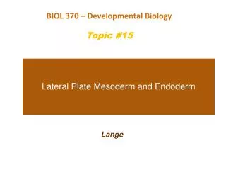
Lateral Plate Mesoderm and Endoderm
760 likes | 2.07k Vues
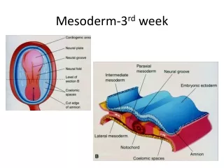
BIOL 370 – Developmental Biology Topic #15. Lateral Plate Mesoderm and Endoderm. Lange. Figure 12.1 Mesodermal development in frog and chick embryos. Notice here how if the chicken is removed from the large yolk (c ), that it will develop in a circular pattern like the frog.

Lateral Plate Mesoderm and Endoderm
E N D
Presentation Transcript
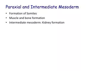
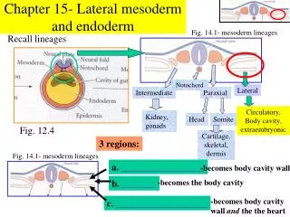
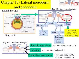
BIOL 370 – Developmental Biology Topic #15 Lateral Plate Mesoderm and Endoderm Lange
Figure 12.1 Mesodermal development in frog and chick embryos Notice here how if the chicken is removed from the large yolk (c ), that it will develop in a circular pattern like the frog.
Figure 12.2 Overview of heart development Heart development comparing chick and mouse.
Heart field formation is a major step in heart development. The first heart field forms a primary aspect of the left ventricle, whereas the second heart field forms the primary portions of the right ventricle and the two atria in organisms with a four chambered heart.
Figure 12.3 Model of inductive interactions involving the BMP and Wnt pathways that form the boundaries of the cardiogenic mesoderm In this model, please note how the lateral plate mesoderm has two regions with two different, but related outcomes. The anterior will become the heart whereas the posterior will become the blood & vessels. Also notice how Wnt, Noggin, and BMP affect development.
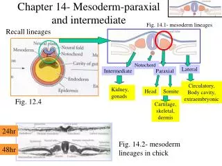
Figure 12.4 Formation of chick heart from splanchnic lateral plate mesoderm • The endocardium is the innermost layer of tissue that lines the chambers of the heart. • Its cells are similar to the endothelial cells that line blood vessels. • The endocardium also provides protection to the valves and heart chambers.
Figure 12.5 Migration of heart primordia • In (a) we see what is called cardia bifida which is a period where the embryo has “two” hearts because there is no interconnection (this is a chicken). • (b) & (c) show zebrafish, but (c) is a mutant form called miles apart (for obvious reasons) • (d) & (e) show mouse with (d) showing heart fusion and (e) showing a Foxp4 deficient mouse
MCPs are multipotent cardiovascular progenitors. They develop into: • endocardium - the innermost layer of tissue that lines the chambers of the heart • endothelium - the thin layer of cells that lines the interior surface of blood vessels and lymphatic vessels • smooth muscle • cardiomyocytes (cardiac muscle cells)
Figure 12.8 Cardiac looping and chamber formation (Part 1) Human heart formation and looping occurs during the 3rd to 4th week after fertilization. *** Also notice how the aortic sac tissues at day 21 become both the aortic sac and pulmonary arteries by birth as we talked about in the prior chapter.
Figure 12.8 Cardiac looping and chamber formation (Part 2) • Notice how significant a change has occurred between stage 9 and stage 10 chick embryo (b & c) • (d & e) both show the mouse heart and show through selective staining how specificity of the atria and ventricles have already occurred (due to differences in their myosin proteins)
Figure 12.9 Formation of the chambers and valves of the heart
Three examples of congenital heart defects Narrowed aorta Occurs in about 1 in every 500 births Occurs in about 1 in every 1500 births Occurs in about 1 in every 2000 births (a) Ventricular septal defect. The superior part of the inter- ventricular septum fails to form; thus, blood mixes between the two ventricles, but because the left ventricle is stronger, more blood is shunted from left to right. (b) Coarctation of the aorta. A part of the aorta is narrowed, increasing the workload on the left ventricle. (c) Tetralogy of Fallot. Multiple defects (tetra = four): Pulmonary trunk too narrow and pulmonary valve stenosed, resulting in a hypertrophied right ventricle; ventricular septal defect; aorta opens from both ventricles; wall of right ventricle thickened from overwork.
Figure 12.10 Embryonic circulatory systems • Lungs and intestines are not functional in the adult sense at embryonic stages. Placental nourishment (including oxygen) necessitate some interesting differences: • Vitelline veins serve to bring nourshment in shell encased eggs (like in birds and reptiles) • Placental veins (umbilical vein) brings nourishment in mammals • The allantoic artery in shell encased bird and reptile eggs will carry wastes away from the embryo • Placental artery (umbilical artery) carries wastes away from the embryo in mammals
Levels of protein structure. Amino acid Amino acid Amino acid Amino acid Amino acid (a) Primary structure: The sequence of amino acids forms the polypeptide chain. (b) Secondary structure: The primary chain forms spirals (-helices) and sheets (-sheets). -Helix: The primary chain is coiled to form a spiral structure, which is stabilized by hydrogen bonds. -Sheet: The primary chain “zig-zags” back and forth forming a “pleated” sheet. Adjacent strands are held together by hydrogen bonds. (c) Tertiary structure: Superimposed on secondary structure. -Helices and/or -sheets are folded up to form a compact globular molecule held together by intramolecular bonds. Tertiary structure of prealbumin (transthyretin), a protein that transports the thyroid hormone thyroxine in serum and cerebro- spinal fluid. (d) Quaternary structure: Two or more polypeptide chains, each with its own tertiary structure, combine to form a functional protein. Quaternary structure of a functional prealbumin molecule. Two identical prealbumin subunits join head to tail to form the dimer.
An example of the progression in complexity of structure in proteins with the final quaternary structure being that of hemoglobin.
Figure 12.11 Adult and fetal hemoglobin molecules differ in their globin subunits When you examine fetal hemoglobin, you see that the protein differences (fetal has the y form and the adult has the B form) lead the fetal RBCs to have a higher oxygen saturation at any state of oxygen pressure. Why is this of value?
Note that BPG (bisphosphoglycergic acid) is a molecule which is lower in fetal hemoglobin than in materinal hemoglobin. This lower interaction gives the higher affinity for fetal hemoglobin.
Figure 12.12 Redirection of human blood flow at birth • The ductusarteriosus is squeezed shut due to pressure changes from the expanding lungs • The embryological foramen ovale (not to be confused with the one in the adult skull) of the heart also closes due to the pressure changes.
Figure 12.13 Aortic arches of the human embryo Aortic arches at day 29 further develop into the leading arteries associated with the aorta and by day 56.
The lymphatic system Regional lymph nodes: Entrance of right lymphatic duct into right subclavian vein Cervical nodes Internal jugular vein Entrance of thoracic duct into left subclavian vein Axillary nodes Thoracic duct Aorta Cisterna chyli Lymphatic collecting vessels Inguinal nodes (a)
Distribution and special structural features of lymphatic capillaries Loose connective tissue around capillaries Venous system Arterial system Venule Arteriole Heart Lymph duct Lymph trunk Lymph node Lymphatic system Lymphatic collecting vessels, with valves Lymphatic capillary Tissue fluid Blood capillaries Lymphatic capillary Tissue cell (a) Filaments anchored to connective tissue Blood capillaries Endothelial cell Flaplike minivalve Fibroblast in loose connective tissue (b)
Figure 12.14 Vasculogenesis and angiogenesis • Vasculogeneis development of new (oiginal) blood vessels • Angiogeneisis the process through which new blood vessels form from pre-existing vessels
Her most famous work: “Studies on the origin of blood vessels and of red corpuscles as seen in the living blastoderm of the chick during the second day of incubation” Journal: Contributions to Embryology Volume 9, 213-262, 1920 Florence Rena Sabin – much of her work involved the discovery of the pathways of blood vessel and lymphatic vessel genesis
Figure 12.15 Vasculogenesis (Part 1) Vasculogenesis – notice how there is blood island formation where primitive blood cells are being made which are derived from undifferentiated mechenchyme. This is very different than in RBC production in the neonate or adult.
Figure 12.16 The lumen, or central space, in the vascular tubes is formed by the fusion of intracellular vacuoles • Lumen formation in vessels: • Vacuole accumulation occurs in lumen cells (endocytosis) • Vacuoles merge forming “megavacuoles” • Continued fusion leads eventually to the lumen being formed.
Figure 12.17 VEGF and its receptors in mouse embryos The VEGF mutant lacks blood vessel development with the yolk sac, and therefore is miscarried.
Figure 12.18 Roles of ephrin and Eph receptors during angiogenesis Eph receptors predominate venous vessels, whereas ephrin-b2 receptors predominate arterial vessels. To encourage angiogenic modeling of capillaries, there is some (as yet unclear) ephrin/Eph interaction that allows formation of these capillary connections.
Figure 12.20 Blood vessel formation in the chick blastoderm One theory explaining blood vessel formation involves the use of VEGF (vascular endothelial growth factor) which may occur as a gradient promoting vessel formation in areas with higher concentration.
Figure 12.22 VEGF-C is critical for the formation of lymphatic vessels A “C” form of VEGF (VEGF-C) is the growth factor for the formation of lymph vessels. Notice the pronounced edema in the mouse embryo that is VEGF-C deficient.
Figure 12.23 Sources of blood cells to adult bone marrow Hematopoiesis – the formation of blood cells is primarily in the red bone marrow in the adult, but embryologically it can occur in diverse places including the liver, the yolk sac and placenta. Hematopoietic stem cells near and around osteoblast cells can differentiate into various blood cell components.
Figure 12.24 A model for the origin of mammalian blood and lymphoid cells
Erythropoiesis: genesis of red blood cells Stem cell Committed cell Developmental pathway Phase 1 Ribosome synthesis Phase 2 Hemoglobin accumulation Phase 3 Ejection of nucleus Early erythroblast Late erythroblast Hemocytoblast Proerythroblast Normoblast Reticulocyte Erythrocyte
Leukocyte formation Hemocytoblast Stem cells Lymphoid stem cell Myeloid stem cell Myeloblast Myeloblast Myeloblast Lymphoblast Committed cells Promyelocyte Promyelocyte Promyelocyte Promonocyte Prolymphocyte Develop- mental pathway Eosinophilic myelocyte Basophilic myelocyte Neutrophilic myelocyte Eosinophilic band cells Neutrophilic band cells Basophilic band cells Monocytes Eosinophils Basophils Neutrophils Lymphocytes (a) (b) (c) (d) (e) Agranular leukocytes Some become Granular leukocytes Some become Plasma cells Macrophages (tissues)
Genesis of platelets Stem cell Developmental pathway Hemocytoblast Megakaryoblast Promegakaryocyte Megakaryocyte Platelets
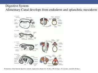
Figure 12.26 Endodermal folding during early human development Notice the differential cuts and how they show organization of the structures that can be visualized in 3D.
Figure 12.27 Formation of glandular primordia from the pharyngeal pouches • Notice how pharyngeal arches develop into: • 1 tympanic cavity • 2 tonsils • 3 parathyroid gland (dorsal) and (ventral) the thymus • 4 more parathyroid gland. • Note that the thyroid is not part of any pharyngeal arch.
Figure 12.28 Regional specification of the gut endoderm and splanchnic mesoderm through reciprocal interactions (Part 1) • cSox2 - • Pdx1 pancreatic and duodenal homeobox 1 • Hox homeobox • cdxC caudal gene C • cdxA caudal gene A
Figure 12.33 Partitioning of the foregut into the esophagus and respiratory diverticulum during the third and fourth weeks of human gestation
Figure 12.34 Wnt signaling is critical for separation of the trachea and early differentiation of the lung
Figure 12.35 The immune system relays a signal from the embryonic lung