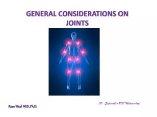
GENERAL CONSIDERATIONS ON JOINTS
GENERAL CONSIDERATIONS ON JOINTS . 30. September 2011 Wednesday. Kaan Yücel M.D., Ph.D . Joints (articulations) are unions or junctions between two or more bones or rigid parts of the skeleton.

GENERAL CONSIDERATIONS ON JOINTS
E N D
Presentation Transcript
GENERAL CONSIDERATIONS ON JOINTS 30. September2011Wednesday Kaan Yücel M.D., Ph.D.
Joints (articulations) are unions or junctions between two or more bones or rigid parts of the skeleton. • Joints exhibit a variety of forms and functions. It is the fact that, whether or not movement occurs between them, it is still called a joint. • Some joints have no movement, others allow only slight movement, and some are freely movable
Classification of Joints Joints are classified according to the tissues that lie between the bones: Fibrous joints Cartilaginous joints Synovial joints
Fibrous joints • The bones are united by fibrous tissue. • The amount of movement occurring at a fibrous joint depends in most cases on the length of the fibers uniting the articulating bones. • The sutures of the cranium are examples of fibrous joints.
A syndesmosis type of fibrous joint unites the bones with a sheet of fibrous tissue, either a ligament or a fibrous membrane. • Consequently, this type of joint is partially movable. • The interosseous membrane in the forearm is a sheet of fibrous tissue that joins the radius and ulna in a syndesmosis.
A dentoalveolarsyndesmosis (gomphosis or socket)is a fibrous joint in which a peglike process fits into a socket articulation between the root of the tooth and the alveolar process of the jaw. • Mobility of this joint (a loose tooth) indicates a pathological state affecting the supporting tissues of the tooth.
Cartilaginous joints The bones are united by hyaline cartilage or fibrocartilage. In primary cartilaginous joints, or synchondroses, the bones are united by hyaline cartilage, which permits slight bending during early life.
Secondary cartilaginous joints, or symphyses, are strong, slightly movable joints united by fibrocartilage. The fibrocartilaginous intervertebral discs between the vertebrae consist of binding connective tissue that joins the vertebrae together.
Synovial joints • The bones are united by a joint (articular) capsule (composed of an outer fibrous layer lined by a serous synovial membrane) spanning and enclosing an articular cavity. • Synovial joints are the most common type of joints and provide free movement between the bones they join. • They are joints of locomotion, typical of nearly all limb joints.
This type of joints has three common features: • Joint cavity: The joint cavity of a synovial joint, like the knee, is a potential space that contains a small amount of lubricating synovial fluid, secreted by the synovial membrane. • Articular cartilage:The articular surfaces are covered by hyaline cartilage
Articular capsule: This structure surrounds the joint and formed of two layers. Inside the capsule, articular cartilage covers the articulating surfaces of the bones; all other internal surfaces are covered by synovial membrane. • Fibrous capsule • Synovial membrane Some synovial joints have other distinguishing features, such as a fibrocartilaginous articular disc or meniscus, which are present when the articulating surfaces of the bones are incongruous.
Ligaments • A ligament is a cordorband of connectivetissueunitingtwostructures. • Articularcapsulesareusuallystrengthenedbyarticularligaments. • Thesearefrom dense connectivetissueandtheyconnectthearticulatingbonestoeachother. • Articularligaments limit theundesired • and/orexcessivemovements of • thejoints.
Articulardisc: Help toholdthebonestogether. Labrum: A fibrocartilaginous ring whichdeepensthearticularsurfaceforone of thebones.
Bursa • Bursae are flattenedsacsthatcontainsynovialfluidtoreducefriction. • Its walls are separated by a film of viscous fluid. • Bursae are found wherever tendons rub against bones, ligaments, or other tendons.
Stability of Joints depends on four main factors: Negativepressure within the joint cavity Shape, size, and arrangement of the articular surfaces Ligaments Tone of the muscles around the joint
Jointvasculatureandinnvervation • Joints receive blood from articular arteries that arise from the vessels around the joint. • Articular veins are communicating veins that accompany arteries (L. venae comitantes) and, like the arteries, are located in the joint capsule, mostly in the synovial membrane. • Joints have a rich nerve supply provided by articular nerves with sensory nerve endings in the joint capsule.
Clinical Notes- JOINTS • Examination of Joints • When examining a patient, the clinician should assess the normal range of movement of all joints. • When the bones of a joint are no longer in their normal anatomic relationship with one another, then the joint is said to be dislocated. Examination of theshoulderjoint
Clinical Notes- JOINTS • Dislocation of Joints • Some joints are particularly susceptible to dislocation because of: • lack of support by ligaments • the poor shape of the articular surfaces, • the absence of adequate muscular support. • The shoulder joint, temporomandibular joint, &acromioclavicular joints are good examples.
Clinical Notes- JOINTS Dislocation of the hip is usually congenital, being caused by inadequate development of the socket that normally holds the head of the femur firmly in position.
Clinical Notes- JOINTS • Damage to Ligaments • Joint ligaments are very prone to excessive stretching and even tearing and rupture. • If possible, the apposing damaged surfaces of the ligament are brought together by positioning and immobilizing the joint. • In severe injuries, surgical approximation of the cut ends may be required.
Clinical Notes- JOINTS Trauma and Infection of Bursae and Tendon Sheaths Bursae and synovial sheaths are commonly the site of traumatic or infectious disease. For example, the extensor tendon sheaths of the hand may become inflamed after excessive or unaccustomed use. An inflammation of the prepatellar bursa may occur as the result of trauma from repeated kneeling on a hard surface.
Clinical Notes- JOINTS • Osteoarthritis • Synovial joints are well designed to withstand wear, but heavy use over several years can cause degenerative changes. • Some destruction is inevitable during such activities as jogging, which wears away the articular cartilages and sometimes erodes the underlying articulating surfaces of the bones.
Clinical Notes- JOINTS Osteoarthritis The normal aging of articular cartilage begins early in adult life and progresses slowly thereafter, occurring on the ends of the articulating bones, particularly those of the hip, knee, vertebral column, and hands.
Clinical Notes- JOINTS • Degenerative joint disease or osteoarthritisis often accompanied by stiffness, discomfort, and pain. • Osteoarthritis is common in older people and usually affects joints that support the weight of their bodies (e.g., the hips and knees).
JOINTS IN THE HEAD • The temporomandibular joint(TMJ) is a synovial joint, permitting gliding and a small degree of rotation in addition to flexion (elevation) and extension (depression) movements. • The bony articular surfaces involved are the mandibular fossa and articular tubercle of the temporal bone superiorly, and the head of the mandible inferiorly. • The two bony articular surfaces are completely separated by intervening fibrocartilage, the articular disc of the TMJ.
JOINTS OF THE VERTEBRAL COLUMN • The vertebral column in an adult typically consists of 33 vertebrae arranged in five regions: 7 cervical, 12 thoracic, 5 lumbar, 5 sacral, and 4 coccygeal. • The joints of the vertebral column include the: • Joints of the vertebral bodies • Joints of the vertebral arches • Craniovertebral(atlanto-axial and atlanto-occipital) joints • Costovertebraljoints • Sacroiliac joints
The joints of the vertebral bodies are designed for weight-bearing and strength. • The articulating surfaces of adjacent vertebrae are connected by intervertebral (IV) discs and ligaments. The IV discs provide strong attachments between the vertebral bodies.
The anulusfibrosus(L. anus, a ring) is a bulging fibrous ring forming the circumference of the IV disc. • The anterior longitudinal ligamentis a strong, broad fibrous band that covers and connects the anterolateral aspects of the vertebral bodies and IV discs. • The posterior longitudinal ligament runs within the vertebral canal along the posterior aspect of the vertebral bodies.
The joints of the vertebral archesare joints between the superior and inferior articular processes of adjacent vertebrae. • Each joint is surrounded by a thin joint capsule. • The laminae of adjacent vertebral arches are joined by broad, pale yellow bands of elastic tissue called the ligamentaflava (L. flavus, yellow).
Therearetwosets of craniovertebraljoints: atlanto-occipitaljoints, formedbetweenthe atlas (C1 vertebra), andtheoccipital bone of thecranium atlanto-axialjoints, formedbetweenthe atlas andaxis (C2 vertebra). Theirdesigngives a widerrange of movementthan in the rest of thevertebralcolumn.
JOINTS OF THE UPPER LIMB Thesternoclavicularjoint (SC) is a synovialjoint. The sternal end of the clavicle articulates with the manubrium and the 1st costal cartilage. The articular surfaces are covered with fibrocartilage. The SC joint is dividedintotwocompartmentsby an articulardisc. TheSC joint is theonlyarticulationbetweentheupperlimbandtheaxialskeleton.
The acromioclavicular joint (AC joint) is a synovial joint, which is formed by the lateral part of the acromion. • The acromial end of the clavicle articulates with the acromion of the scapula. • The articular surfaces, covered with fibrocartilage, are separated by an incomplete wedge-shaped articular disc. • The acromion of the scapula rotates on the acromial end of the clavicle.
The glenohumeral (shoulder) joint is a synovial joint that permits a wide range of movement; however, its mobility makes the joint relatively unstable. The large, round humeral head articulates with the relatively shallow glenoid cavity of the scapula, which is deepened slightly but effectively by the ring-like, fibrocartilaginousglenoid labrum (L., lip).
The glenohumeral joint has more freedom of movement than any other joint in the body. • This freedom results from the laxity of its joint capsule and the large size of the humeral head compared with the small size of the glenoid cavity.
The elbow joint, a synovial joint, is located inferior to the epicondyles of the humerus. • Thereare humeroulnar and humeroradial articulations. • The collateral ligaments of the elbow joint are strong triangular bands that are medial and lateral thickenings of the fibrous layer of the joint capsule. • The radial collateral ligament • The ulnar collateral ligament • Flexion and extension occur at the elbow joint. • Intratendinous olecranon bursa • Subtendinous olecranon bursa • Subcutaneous olecranon bursa
The proximal (superior) radio-ulnar joint is a synovial joint that allows movement of the head of the radius on the ulna. The radial head is held in position by the anular ligament of the radius. The distal (inferior) radio-ulnar joint is a synovial joint. The radius moves around the relatively fixed distal end of the ulna.
The wrist (radiocarpal) jointis a synovial joint. The ulna does not participate in the wrist joint. • The distal end of the radius and the articular disc of the distal radio-ulnar joint articulate with the proximal row of carpal bones, except for the pisiform. • Theintercarpal (IC) jointsinterconnect the carpal bones. • The carpometacarpal (CMC), intermetacarpal (IM) joints, metacarpophalangeal joints, and interphalangeal joints are other joints in the hand.
JOINTS OF THE PELVIS Pubicsymphysisfibrocartilaginousconsists of an interpubicdiscandsurroundingligamentsunitingthebodies of thepubicbones in themedianplane. Lumbosacraljoints; L5 and S1 vertebrae articulate The other joint in the pelvis is the sacrococcygeal joint.
JOINTS OF THE LOWER LIMB The joints of the lower limb include the articulations of the pelvic girdle—lumbosacral joints, sacroiliac joints, and pubic symphysis.
The hip joint forms the connection between the lower limb and the pelvic girdle. It is a strong and stable synovial joint. • The head of the femur is the ball, and the acetabulum is the socket. • The hip joint is designed for stability over a wide range of movement. • Next to the glenohumeral (shoulder) joint, it is the most movable of all joints. • During standing, the entire weight of the upper body is transmitted through the hip bones to the heads and necks of the femurs.
The round head of the femur articulates with the cup-like acetabulum of the hip bone. • The lip-shaped acetabular labrum (L. labrum, lip) is a fibrocartilaginous rim attached to the margin of the acetabulum, increasing the acetabular articular area by nearly 10%. • The hip joints are enclosed within strong joint capsules, formed of a loose external fibrous layer (fibrous capsule) and an internal synovial membrane.