Download

1 / 33
680 likes | 1.74k Vues
Anatomy of swallowing. General considerations. Series of activities that occur within a matter of seconds. Traditionally described as a reflex, the process is more properly regarded as a programmed motor behaviour .

E N D
General considerations • Series of activities that occur within a matter of seconds. • Traditionally described as a reflex, the process is more properly regarded as a programmed motor behaviour. • Swallowing is initiated when food or liquid stimulates sensory nerves in the oropharynx.
In a 24-hour period, an average person will swallow between 600 and 1000 times • Only some 150 will relate to feeding; the remainder occur to clear continuously produced saliva and are less frequent at night.
Eating and drinking are basic human pleasures • Problems associated with swallowing can impact dramatically upon the quality of life. • Swallowing disorders are usually symptoms of other complex diseases • An inability to swallow may adversely affect nutritional status and therefore indirectly exacerbate the underlying disease
Aside from the risk of asphyxiation through choking, swallowing disorders can also be a direct cause of morbidity and mortality as a result of aspiration of food, liquid or possibly refluxed gastric acid contents, causing bacterial infection or tissue damage.
For descriptive purposes, the process has been divided into four phases: • oral preparatory • oral transit/transfer • pharyngeal • oesophageal. • The boundaries between these phases are not entirely clear, thus, for example, the demarcation between the first and second phases is defined primarily by convention
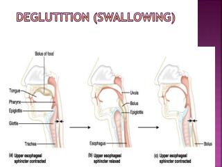
Food is reduced in the mouth to a consistency suitable for swallowing and is formed into a cohesive bolus (oral preparatory phase). • The bolus is delivered to the oropharynx (oral transit/transfer phase), transported down the pharynx past the airways and through the upper oesophageal sphincter (pharyngeal phase), and then transported down the oesophagus to the stomach (oesophageal phase).
The oral preparatory and oral transit/transfer phases are voluntary and under cortical control, whereas the pharyngeal and oesophagealphases are involuntary and controlled by the brainstem. • Airway protection is vital during the pharyngeal phase.
ORAL PREPARATORY PHASE • Chewing • Food reduced by the grinding action of the teeth and simultaneously mixed with saliva • lips are maintained as a tight l seal by the contraction of orbicularisoris • Buccinator performs a similar function for the cheeks. • Vestibule normally remains empty, and any food that enters the vestibule is returned to the oral cavity proper.
Buccinator also keeps the cheeks taut, ensuring that they are kept clear of the occlusal surfaces of the teeth during chewing • Loss of the nerve supply to buccinatorresults in painful and repeated lacerations of the cheeks.
The soft palate is depressed during this phase and premature spillage of food is common. • Spillage occurs because the soft palate is not in continuous contact with the posterior part of the tongue
Bolus formation appears to involve several cycles of food being transported from the anterior to the posterior part of the tongue through the palatoglossal and palatopharyngeal arches until a bolus accumulates on the oropharyngeal surface of the tongue (retrolingual loading).
Throughout this phase, the lateral and rotatory tongue movements are crucial for normal bolus formation. • If effective tongue movements do not occur, chewing will be compromised. • End of this phase marked by the tongue holding the bolus of food that has been formed against the hard palate in readiness for transport to the posterior part of the oral cavity.
ORAL TRANSIT/TRANSFER PHASE • Bolus finally transported through the palatoglossal and palatopharyngeal arches into the oropharynx. • Genioglossus raises both the tongue tip and the part of the tongue immediately behind the tip. • The soft palate is then fully lowered by contraction of palatoglossus and palatopharyngeus, and the posterior part of the tongue is simultaneously elevated:
The apposed soft palate and tongue form a tight seal that helps to prevent premature entry of the bolus into the pharynx. • Orbicularisoris and buccinator remain contracted, so keeping the lips and cheeks taut and the bolus of food central in the oral cavity.
Bolus is accommodated in a shallow midline gutter that forms along the dorsum of the tongue, probably as a result of the co-contraction of the styloglossi and the genioglossi, aided by the superior longitudinal and transverse fibres of the intrinsic muscles.
Mandible is elevated and the mouth is closed. • Floor of the mouth and the anterior and middle portions of the tongue are elevated, by co-contraction of the suprahyoid group of muscles (mylohyoid, digastric, geniohyoid and stylohyoid
Effectiveness of the suprahyoid muscles is increased as they contract against a fixed mandible (the mouth does not have to be closed to swallow, but it is much harder to swallow if it is open). • Contraction of stylohyoid elevates the more posterior parts of the tongue and empties the longitudinal gutter. At the same time, the tongue flattens, probably as a result of the contraction of hyoglossus and some of the intrinsic lingual muscles, especially the vertical fibres.
At the same time, the tongue flattens, probably as a result of the contraction of hyoglossus and some of the intrinsic lingual muscles, especially the vertical fibres.
The elevated, flattened tongue pushes the bolus against the hard palate, and the sides of the tongue seal against the maxillary alveolar processes, helping to move the bolus further posteriorly. • Contraction of styloglossus and mylohyoid completes the elevation of the posterior part of the tongue.
At the same time, the posterior oral seal relaxes and the posterior tongue moves forward • the overall effect is sweeping or squeezing the bolus towards the pillars of the fauces, finally delivering it to the oropharynx where the pharyngeal aperture is initially increased and then closed.
PHARYNGEAL PHASE • involuntary and the most critical • involves the pharynx changing from being an air channel (between the posterior nares and laryngeal inlet) to a food channel (from the fauces to the upper end of the oesophagus).
The airway is protected from aspiration during swallowing by elevation of hyoid and larynx. and by resetting respiratory rhythm • Airflow ceases briefly as the bolus passes through the hypopharynx • The total time that elapses from the bolus triggering the pharyngeal phase to the re-establishment of the airway is barely 1 second.
Nasopharynx is sealed off from the oropharynx by activation of the superior pharyngeal constrictor and contraction of a subset of palatopharyngealfibres to form a variable, ridge-like structure (Passavant's ridge) against which the soft palate is elevated.
the pharyngeal ridge becomes hypertrophic in an infant with a cleft palate, presumably in an attempt to produce a seal to the nasal airway. • Ineffective velopharyngeal closure may result in nasal regurgitation of food.
The airway is sealed at the laryngeal inlet by closure of the glottis • Epiglottis is retroflexed over the laryngeal aditus as a result of passive pressure from the base of the tongue and active contraction of the aryepiglottic muscles.
hyoid bone and larynx are raised and pulled anteriorly by the suprahyoid muscles and the longitudinal muscles of the pharynx • laryngeal inlet is brought forward under the bulge of the posterior tongue, i.e. out of the path of the bolus.
This action helps expand the hypopharyngeal space and relax the upper oesophageal sphincter, which is also raised by several centimetres. • The bolus passes over the reflected anterior surface of the epiglottis and is swept through the laryngopharynx to the upper oesophageal sphincter.
This action helps expand the hypopharyngeal space and relax the upper oesophageal sphincter, which is also raised by several centimetres. • The bolus passes over the reflected anterior surface of the epiglottis and is swept through the laryngopharynx to the upper oesophageal sphincter.
The tongue driving force, or the tongue thrust pressure force, is a positive pressure that squeezes the bolus towards the laryngopharynx. • It is generated by the upward movement of the tongue pressing the bolus against the contracting pharyngeal wall and requires a tight nasopharyngeal seal (created by elevation of the soft palate).
The pharyngeal constrictors generate a positive pressure wave behind the bolus. • Their sequential contraction may facilitate clearance of the pharyngeal walls
OESOPHAGEAL PHASE • Begins after the relaxation of the upper oesophageal sphincter has allowed the bolus to enter the oesophagus. • Sequential waves of contractions of the oesophageal musculature subsequently propel the bolus down to the lower oesophageal sphincter, which opens momentarily to admit the bolus to the stomach
The oesophageal phase of swallowing is much more variable than the other phases, and lasts between 8 and 20 seconds.