Cleavage site
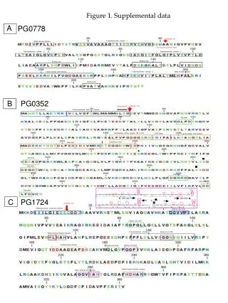
Figure 1. Supplemental data. A. PG0778. Cleavage site. Transmembrane domain -1. Metal bindiing domain. Twin arginine peptide signal. Transmembrane domain -2. Transmembrane domain -3. Binding site. Binding site. Lectin binding CHO domain. Transmembrane domain -4.

Cleavage site
E N D
Presentation Transcript
Figure 1. Supplemental data A PG0778 Cleavage site Transmembrane domain -1 Metal bindiing domain Twin arginine peptide signal Transmembrane domain -2 Transmembrane domain -3 Binding site Binding site Lectin binding CHO domain Transmembrane domain -4 Metal binding domain Binding site B PG0352 Cleavage site Signal peptide Inner domain Transmembrane helix domain Asp motif Exosialidase activity domain 182 to 240 FRIP region Lectin binding CHO domain ‡ Consensus motif Asp motif ▲ ▄ Asp motif ◙ Asp motif Asp motif Asp motif Asp motif ‡ ▲ ▄ 230-240 290-300 C PG1724 360-370 ◙ 410-420 Cleavage site Metalloprotease consensus domain Metalloprotease consensus domain Glycine rich motif Glycine rich motif Glycine rich motif Transmembrane domain Metal binding Histidine domain Endoprotease domain Glycine rich motif Glycine rich motif Metal binding Histidine domain Lectin binding CHO domain
Figure 1.supplemental data. Amino acid sequences of sialidase/O-sialoglycoprotease genes of P. gingivalis showing various unique domains and motifs. • Amino acid sequence of PG0778 showing 4 transmembrane domains, two metal binding domains and one lectin binding carbohydrate domain. There is a twin arginine peptide signal at the 39th position coinciding with the cleavage and specific binding sites. • B. Amino acid sequence of PG0352 showing an inner domain, transmembrane domain and an outer domain. There is a specific exonuclease activity domain consisting of a FRIP region, a non catalytic lectin binding domain and a consensus Asp box domain spanning amino acid positions 182 to 240. The Asp box sequences in the protein are shown in an inset box with positions of their location. Non-specific DxxD (aspartic acid motifs) are noted in 7 positions. The transmembrane helix domain consists of signal peptide sequence and a cleavage site. • C. Amino acid sequence of PG1724 showing N terminal metalloprotease consensus domain bearing a cleavage site. Metal binding histidine domains are indicated at positions 113 and 290. A transmembrane domain spans position 136 to 152. Glycine rich motifs were found distributed throughout the protein. A lectin binding domain is indicated at positions 277 – 282.
Figure 2. Supplemental data. 482 Actinomyces naeslundii (ANA1493) Clostridium perfringens Tannerella forsythia (TF0035) Porphyromonas gingivalis (PG0352) P.gingivalis(33277)(PGN1608) Actinomyces naeslundii (ANA2709) Bacteroides fragilis 785 Tannerella forsythia (TN2207) 867 CONSENSUS Actinomyces naeslundii (ANA1493) Clostridium perfringens Tannerella forsythia (TF0035) Porphyromonas gingivalis (PG0352) P.gingivalis (33277)(PGN1608) Actinomyces naeslundii (ANA2709) Bacteroides fragilis Tannerella forsythia (TN2207) CONSENSUS ▲ Figure 2. supplemental data. Consensus amino acid signature sequence of sialidases among oral pathogens. Consensus amino acid signature sequence of sialidase Asp box (SxDxGxTW) found in PG0352 and the other members of Clad A (Fig. 1).
Figure 3. Supplemental data Identification of Asp box like motifs and a consensus signature motif-DAxG/DAxD in O-Sialoglycoprotease clusture
Figure 3.supplemental data Identification of Asp box like motifs(shown in black under score) and consensus sequence motifs of DAxG /DAxD (shown in blue under score) present in the O-sialoglycoprotease clustures.
Figure 4. Supplemental data. A C B Model of PG0352 -sialidase Model of PG778 O-sialoglycoprotease Model of PG1724 O-sialoglyco protease 2 1 1 2 90o 90o 60o 4 3 3 4 4 90o 90o 60o Terminal clef and molecular groove for optimal interaction. The model shows more hydrophilic regions suggestive of more active sites. loose confirmation suggestive of weak interactions sites with less hydrophilic regions Compact configuration showing less hydrophilic regions 1 2 4 3
Figure 4.supplemental data. In silico protein modeling • Protein model of PG0778 showing terminal clef and molecular groove for optimal interaction. The model shows more hydrophilic regions suggestive of more active sites than the other O-sialoglycoprotease PG1724. The ribbon model showing the sheets forming end propeller and flanking helixes (1). The surface structure of the protein showing an interactive tunnel coinciding with the terminal cleft was shown.The hydrophilic areas in the protein are colored red (2-4). • B. Protein model of PG0352 showing a compact configuration with less hydrophilic • regions. The model shows the sialidase protein is a monomer containing six sheets (1 and 2). The protein surface show less hydrophilic regions (3 and 4). • C. Protein model of PG1724 showing end propeller structure (1 and 2) with a loose • confirmation with less hydrophilic regions on the surface of the protein (3 and 4).
Figure 5. Supplemental data Assay of P.gingivalis complemented mutants showing restored defects A B C Figure 5.supplemental data A. Graph showing the total protease and sialidase activity of the P.gingivalis complemented mutants. B. Graph showing the gingipain activity of the P.gingivalis complemented mutants C. Graph showing the substrate utilisation of the P.gingivalis complemented mutants