Foot & Ankle
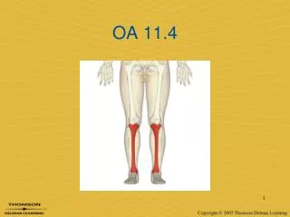
Foot & Ankle. Anatomy. Anatomy - Medial. Anatomy - Lateral. Talocrural Joint. Subtalar Pronation – Closed Chain. Subtalar Pronation – Closed Chain. Subtalar Supination – Closed Chain. Subtalar Pronation & Supination Model – Closed Chain. Gait Review.

Foot & Ankle
E N D
Presentation Transcript
Transverse Tarsal/Midtarsal/Chopart’s Joint Talonavicular Joint Calcaneocuboid Joint
Midtarsal Joint Pronation • STJ unlocks MTJ Supination • STJ locks up MTJ
Midtarsal Joint Motion - Closed Chain Pronation Neutral Supination
Abnormal Biomechanics • Breakdown of CT • Reduced muscle efficiency • Change in muscle function • Poor alignment – Osseous Deformity • Dysfunction and Pathology • Reduced ability to attenuate GRFs
Pronation Closed Chain • Calcaneus eversion (valgus) • Talus adduction (IR - vertical axis) • Talus plantarflexion • Tibial IR
Normal Pronation in Gait Normal Range: • 6 - 100 Excessive: • 130+
Abormal Pronation in Gait • Excessive in magnitude • Excessive in duration • Occurs at wrong time Causes: • Intrinsic deformities • Extrinsic deformities
RF/Subtalar Joint Varus • Inversion deformity of calcaneus • No change in relationship of RF on FF Etiology • Congenital/developmental • failure of talus to derotate
Forefoot Varus • Most Common • Insufficiency of 1st ray • Dorsiflexed/hypermobile 1st ray • Congenital deformity • Inversion of forefoot (metatarsals) relative to rearfoot in STJ neutral
Forefoot Varus (Compensated) - Pathomechanics • During WA - excessive pronation to get 1st ray on ground • Max. pronation occurs @ HO • Pronation remains thru propulsion • Foot never becomes rigid lever • Instability
Forefoot Varus - Compensation • Prolonged / excessive pronation • Calcaneal valgus • Unlocking of forefoot during propulsion • Insufficient pulley system
Forefoot Varus - Pathology • Hypermobile 1st ray • Excessive forces on 2nd MET • Prolonged / excessive tibial torsion and/or IR • Excessive anteversion of hip
Forefoot Varus - Uncompensated • Rigid Foot • Lateral ankle sprains • S.I. Joint Dysfunction • ITB Dysfunction
Subtalar Varus and Compensated Forefoot Varus FF Varus • acquired soft tissue contracture at MTJ • 20 compensatory pronation for a STJ varus
Ankle Joint Equinus • Fixed limitation of DF @ TCJ • < 100 of DF when in STJ neutral and knee / Etiology • tight gastrocnemius • spasticity • flattened dome of talus • Fx, arthritis, trauma
Ankle Joint Equinus - Pathomechanics • pronation 20 to DF • loss of ankle rocker • tibia unable to move anterior to talus tibia and talus move anterior to calcaneus • DF of RF at FF • and prolonged pronation during propulsion
Compensated Ankle Joint Equinus • Excessive STJ pronation • Calcaneal valgus/eversion • Inefficient pulleys • DF of RF on FF
Uncompensated Ankle Joint Equinus • Genu Recurvatum • Early heel rise • Excessive abduction and ER of LE