Download
1 / 9
90 likes | 386 Vues
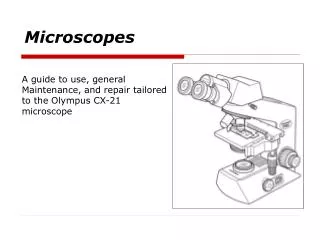
The History of Microscopes: For Dummies. Discovery of the Cell. Robert Hooke (1665) – English scientist who looked at thin slices of cork (dead oak tree bark) under a light microscope and noticed tiny “compartments” that he named cells .

E N D
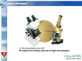
Discovery of the Cell • Robert Hooke (1665) – English scientist who looked at thin slices of cork (dead oak tree bark) under a light microscope and noticed tiny “compartments” that he named cells. • Anton van Leeuwenhoek (1700) – Dutch fabric merchant and amateur scientist developed a better microscope and observed tiny living organisms in pond water, that he named “animalcules.” Hooke’s original drawing of cork under a microscope.
Discovery of the Cell • Matthias Schleiden (1838) - a German botanist who viewed plant parts under a microscope and discovered that plant parts are made of cells. He is considered to be the co-founder of cell theory together with Schwann, with whom he consulted. • Theodor Schwann (1839) - a German biologist who viewed animal parts under a microscope and discovered that animals were made up of cells. He extended Schleiden's cell theory in plants to animals, stating that all living things are composed of cells.
Discovery of the Cell • Rudolph Virchow (1855) - a German physician who stated that all living cellscome only from other living cells. By combining the work of all of the scientist mentioned, the Cell Theory was born…
The Cell Theory – mid 1800s Cell theory is the basis for the way that biologists study living things. Cell Theory is the most basic condition for determining if something is living. • All organisms are made of cells. • Cells are the basic unit of structure and function in living things. • New cells come from pre-existing cells.
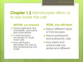
Light Microscopes (LM) These are used to magnify objects up to about 1000 times their actual size (bacteria or larger) Used to examine living or dead specimens Light switch
Most cells are transparent, making them difficult to see without staining them with dyes! Onion cells stained with dye Human cheek cells stained with dye Arterial epithelial cells stained with fluorescent dye
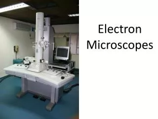
High Power Microscopes! • In the 1950s, electron microscopes were developed which used a beam of electrons instead of a beam of light. • Two Types – only view dead specimens • Scanning Electron Microscope (SEM) • Examines the surface structures of cells • Transmission Electron Microscope (TEM) • Examines the internal structure of cells Micrograph – a photograph of the view through a microscope Red algae (TEM) Human egg (SEM)
Micrograph of a sickle cell RBC Micrograph of RBC Micrograph of a flea Micrograph of head louse Sperm production Surface of a strawberry