Peripheral Nerve Blocks using Nerve Stimulator
750 likes | 2.07k Vues
Peripheral Nerve Blocks using Nerve Stimulator. Dr.D.KANNAN. D.A., D.N.B., Consultant Anaesthesiologist, Meenakshi Mission Hospital And Research Centre, Madurai. Peripheral Nerve Blocks using Nerve Stimulator. Introduction Nerve stimulator Drugs and toxicity Advantages of block Anatomy

Peripheral Nerve Blocks using Nerve Stimulator
E N D
Presentation Transcript
Peripheral Nerve Blocks using Nerve Stimulator Dr.D.KANNAN. D.A., D.N.B., Consultant Anaesthesiologist, Meenakshi Mission Hospital And Research Centre, Madurai.
Peripheral Nerve Blocks using Nerve Stimulator Introduction Nerve stimulator Drugs and toxicity Advantages of block Anatomy Nerve blocks Femoral N Obturator N Sciatic N Saphenous N Ankle block
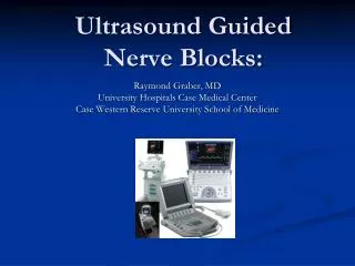
EQUIPMENTS Nerve Stimulator Unipolar needles of varying length Flexible catheter Electrode
Nerve Stimulator Current range from 0.1-5.0 mA Pulse Frequency 1 Hz -Mixed nerve 2 Hz - Sensory nerve
Nerve Stimulator The electrical current required to trigger muscle contractions correlates with the distance of the tip of the needle to the nerve. That means that the closer the needle is to the nerve, the lower the electrical current that is required to induce contractions or sensory responses
Stimulation and Injection tecnique Initial current 2-3 mA Frequency 1-2 Hz Threshold current 0.3- 0.5 mA Aspiration test 5- 10 ml LA injected slowly Increase the current to initial level No stimulatory response - inject the remaining drug Recurring response - May indicate intra vascular needle position
Drugs 2% Plain Lignocaine 3 mg / kg 2% Lignocaine with adrenaline 7 mg / kg of 0.5% Bupivacaine 2 mg / kg o.75 % Ropivacaine 2-3 mg / kg Analgesic: 0.125% Bupivacaine, 0.2% Ropivacaine, Opiods, Clonidine.
Advantages of Block On Arrival block Preemptive analgesia Post operative pain relief Rescuing a risky patients Less complications Cost factor
Lower Limb innervations The innervations of the leg is derived from the lumbar and sacral plexuses Lumbar plexus formed by T12–L4 The main branches are Lateral cutaneous N of thigh Femoral N Genitofemoral N Obturator N
Lower Limb innervations Lumbo sacral plexus formed by L4 –S5 Main branches are Sciatic nerve Posterior cutaneous nerve of thigh Pudental nerve
Lumbosacral Plexus Lateral cutaneous N of thigh Femoral N Genitofemoral N Sciatic N Obturator N Pudental N
Distribution of Lumbo sacral plexus Lateral cutaneous nerve of thigh Femoral nerve Sciatic nerve Obturator nerve
Lower Limb Blocks Femoral Nerve Block Obturator Nerve Block Lateral cutaneous Nerve Block Trans gluteal Sciatic Nerve Block Popliteal Saphenous Nerve Block Ankle Block
Femoral Nerve Block Indications Operative procedures in areas supplying the femoral and lateral femoral cutaneous nerves In combination with proximal sciatic nerve block, operative procedure on the whole leg. Analgesia
Femoral Nerve Block Contraindications No particular Side effects / complications Vessel puncture (of the femoral vein or artery)
Femoral Nerve Block Anatomical landmarks Groin Femoral artery Anterior superior iliac spine Pubic tubercle Inguinal ligament
Femoral Nerve Block Blockade technique The patient lies on his back, his leg loosely abducted and turned to the outside. Puncture site: 2cm caudad to the groin, 1 – 2 cm lateral to the femoral artery.
Femoral Nerve Block Puncture direction: 30 ̊ – 40̊ cranial parallel to the artery. Puncture depth: 2 – 4 cm. Positive stimulatory response from the femoral nerve: Rectus muscle of the thigh (“dancing patella”).
What to do when? Stimulation of the Sartorius muscle (medial contraction) occurs Puncture direction usually too medial. Retract the needle, and shift it slightly to the lateral. Direct stimulation of the Sartorius muscle (rare): Puncture direction is usually too lateral Shift the needle slightly to the medial. Femoral artery puncture: Retract the needle Shift puncture direction to the lateral.
Potential errors and hazards LA injection in the case of Sartorius muscle stimulation. Intravascular injection
Obturator nerve block Indications Suppression of the adductor reflex for the transurethral lateral bladder wall resection. Treatment of adductor spasm. Adjunct to the femoral nerve blocks for postoperative medial knee joint pain. Analgesia.
Obturator nerve block Contraindications No particular Side effect / complications Vessel puncture (obturator artery or vein)
Anatomical landmarks Origin of adductor longus muscle Pubic tubercle Femoral artery Anterior superior iliac spine
Obturator nerve block Blockade technique The patient is supine on his back, his leg is rotated outwardly and abducted. Puncture site: 5 – 10 cm beneath the pubic tubercle directly lateral to the tendon origin of the adductor longus muscle.
Obturator nerve block Puncture direction approx. 45 ̊ craniolateral pointing towards the anterior iliac spine. Puncture depth: 4 – 6 cm. Positive stimulatory response from adductor group.
What to do when? Persistent adductor spasm despite proper ONB Perform additional femoral block Note The adductor reflux for TURP can reliably suppressed by separate Obturator Nerve block Not by Femoral N block nor Spinal anaesthesia
Transgluteal sciatic nerve block Indications Operative procedure in areas supplying the sciatic nerve In combination with psoas compartment block / femoral nerve block for operations on the whole leg Analgesia
sciatic nerve block Contraindications No particular Side effects / complications Vessel puncture (inferior gluteal artery)
Transgluteal sciatic nerve block Anatomical landmarks Greater trochanter Posterior superior iliac spine Ischial tuberosity Sacral hiatus Puncture site
Transgluteal sciatic nerve block Blockade technique The patient is placed in the lateral recumbent position; hip flexed 45 ̊, knee flexed 70 ̊(“Stable recumbent position”)
Puncture site 4 – 5 cm mediocaudal on the mid-perpendicular lines between greater trochanter and posterior superior iliac spine; connecting line between the greater trochanter and sacral hiatus intersects the insertion point at the mid-perpendicular line.
Puncture depth 5 – 8 cm Positive stimulatory response From the peroneal or tibial nerves: Extensors or flexors of the foot or toes Dosage 20-40 ml
What to do when? Contraction of the Gluteus maximus muscle (= direct stimulation) occurs: Continue to advance the needle until the typical response is elicited. Stimulatory response from the ischiocrural muscle group: LA injection possible Delayed onset of action
What to do when? Bone contact, No Stimulatory response: Correct insertion direction to midline between greater trochanter and ischial tuberosity Potential errors and hazards LA injection upon stimulatory response from the gluteal muscles.
Popliteal sciatic nerve block Indications Operation procedure in the area supplying the sciatic nerve of the lower leg and foot In combination with saphenous nerve block, operations on the whole lower extremity. Analgesia.
Popliteal sciatic nerve block Contraindications Stent (relative) Side effects / complications Vessel puncture (popliteal artery/vein)
Popliteal sciatic nerve block Anatomical landmarks Popliteal fossa Popliteal fold Long head of the biceps femoris muscle Medial and lateral epicondyle of the femur
Popliteal sciatic nerve block Blockade technique Patient is either in prone position or lying on his side, leg extended Puncture site 8-12 cm above the fold of popliteal fossa at the medial edge of biceps femoris muscle.
Popliteal sciatic nerve block Puncture depth 2-4 cm Positive stimulatory response From the Peroneal and Tibial nerve (extensors or flexors of the foot or toes) Dosage: 30 – 40 ml
What to do when…? Femur contact occur: Insertion too deep and too medial Retract the needle Correct direction or insertion site to the lateral, reduce insertion depth. Vessel puncture popliteal artery/vein: Puncture too depth and too medial Retract the needle Correct insertion direction to the lateral, reduce insertion depth.
Potential errors and hazards Puncture site is too for caudad (popliteal fold): It may be that the tibial (med.) and perpneal nerve (lat.) are separated so far apart that complete blocked cannot be achieved with a single LA injection at the two sciatic branches.
Saphenous nerve block Indications Operative procedures in the area supplying the saphenous nerve. In combination with distal sciatic nerve block for operations on the whole lower leg and foot. Analgesia.
Saphenous nerve block No contraindications /side effects Anatomical Landmarks Petellar crest Sartorius muscle Vastus medialis muscle
Saphenous nerve block Blockade technique Patient lies on his supine back with extended leg in neutral position, rotated slightly outwards. Puncture site: Approx. 2 – 4 cm cranial and medial of the upper patellar crest over the sartorius muscle. Puncture direction perpendicular through the muscle up to the subsartorial fatty tissue.
Saphenous nerve block Puncture depth: 3 – 5 cm. Positive response include paresthesias on the medial lower leg at a pulse duration of 1.0 ms. Dosage 10 – 15 ml LA
What to do When…? Motor stimulatory response comes from the Vastus medialis muscle Consider as positive Inject the drug Alternative technique Subcutaneous infiltration below the medial knee joint from the medial head of gastronemius muscle to the tibial tuberosity
Ankle Block An ankle block is essentially a block of four branches of the sciatic nerve Deep Peroneal N Superficial Peroneal N Tibial N Sural N one cutaneous branch of the femoral nerve Saphenous N