Download
1 / 30
350 likes | 908 Vues
Regional Anaesthesia An introduction to peripheral nerve blocks. Dr Aasifa Tredray Consultant Anaesthetist Clinical Governance meeting July 11 th 2013. Aim of presentation. Equipment Anatomy of an axillary block Practical aspects of an axillary block Complications.

E N D
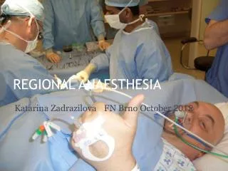
Regional AnaesthesiaAn introduction to peripheral nerve blocks Dr Aasifa Tredray Consultant Anaesthetist Clinical Governance meeting July 11th 2013
Aim of presentation • Equipment • Anatomy of an axillary block • Practical aspects of an axillary block • Complications
Peripheral Nerve Blocks Distribution of local anaesthetic around nerves causing them to become numb and allow surgical procedures to be done. • Advantages patient factors-analgesia, no GA, short starvation times, quicker recovery, long term benefits hospital factors-more efficient lists, more DSU patients, financial savings • Disadvantages patient suitability-contraindications logistics-time, list order, equipment, location Overall high patient satisfaction rate so we should encourage and facilitate its use
Principle of Ultrasound Transducer contains Piezo electric crystals - convert electric signal into sound waves Sound waves are then either absorbed or reflected back as they pass through structures in the body Reflected waves are then convert by machine into a digital 2D image seen on screen High frequency linear probes(5-12MHz) allows higher resolution but poor penetration so good for superficial structures Low frequency curvilinear probes (2-5MHz) will allow greater penetration but poorer resolution but good for deeper structures
Knobology!! Depth Gain Amplification of received ultrasound information which will lighten whole image Noise and artefacts are also amplified so best kept at lowest level that allows clear distinction of desired structures. Latest machines offer complex computer controlled auto- gain buttons
Doppler Describes apparent change in sound frequency as source moves towards or away from probe Away from probe as blue and flow towards probe as red (i.e. not necessarily corresponding to arterial and venous) If Flow is perpendicular to probe then there will be Doppler shift and there will be no coloured flow on screen
Needling • Out of plane • In plane
Peripheral Nerve Stimulators PNS is useful adjunct to USM as it increases safety of block Settings are pulse duration 0.05-0.1ms, frequency 2Hz and current starting at 0.5-1mA Current – distance of needle to nerve and which nerve is being stimulated If twitches seen current is then reduced until no motor response is seen Ideal is for twitches to disappear by 0.2/0.3mA If twitches disappear at a current > 0.5mA usually needle is too far away from nerve, so block will have a very slow onset or be unsuccessful If twitches still present at current < 0.2mA,or increased resistance or pain on injection may suggest intraneural needle placement
Pulse width Pulse width is the duration the current needs to be applied to a nerve to cause a stimulus/contraction Different nerves have different pulse widths Possible to stimulate motor nerves but not sensory or pain nerves by using a short pulse width of 0.05-0.1ms.
Frequency Rate at which the twitches/contractions are seen Ideal comfortable stimulation is 1 to 2 Hz • Higher frequency will give more frequent feedback to operator, but can cause greater discomfort to patient • Not all nerves twitch as demonstrated when using the USM where you can see that your needle is in close proximity to nerve and no motor responses elicited even at currents 1.5mA • Patients with neuropathy do not respond normally to PNS
Common St George’s Blocks • Brachial plexus blocks Interscalene block Supraclavicular and Infraclavicular blocks Axillary block • Nerve blocks at the elbow • Nerve blocks of the lower limb
Axillary Blocks • Anaesthetic pre-operative assessment • Consent • Plan B • Anaesthetic room WHO sign in Full monitoring and 1 BP measurement taken Establish IV access Emergency/anaesthetic drugs available • Position patient with arm out at 90 degrees Axilla and whole arm exposed Make sure they are comfortable
Axillary Blocks Equipment Ultrasound machine – M turbo/Micromax/S nerve 50mm insulated nerve stimulating needle +/- Peripheral Nerve Stimulator ECG and needle connections Sterile gel and sheath for probe • Local Anaesthetic mixture
Set up of block Sit down and get comfortable • Select appropriate probe • US machine set for nerve examination • Use adequate US gel to provide an air free interface • Gel applied to probe, then covered with sterile sheath • Skin cleaned with antiseptic spray -2% chlorhexidene is OK for peripheral blocks but not for spinals or epidurals • Sterile Gel applied to skin • Place probe on patient and orientate image (blue dot) • Assess anatomy and look for key landmarks
White / hyperechoic Black / hypoechoic Nerves close to spinal cord appear hypoechoic Nerves pick up connective tissue as they move away from the spinal cord so change and become hyperechoic
Local Anaesthetic to skin • Remove all air from LA syringe and flush needle- air really distorts the ultrasound image • Connect needle to PNS if being used • Choose entry point with PAJUNK needle • Recheck settings of PNS • When needle is in correct position for nerve, anaesthetist will ask you to ASPIRATE gently then inject 0.5ml- 1ml LA • STOP injection if too hard- high pressure may suggest intra neural placement • We will assess spread of LA- if good will ask you to repeat • Ideal is for spread to surround entire nerve
Step by Step procedure Axillary Block Post block- keep monitoring patient and cycle BP every 3-5mins Don’t rush and allow time for it to work Assess numbness using cold spray-looking for a temperature difference. Keep questions simple with yes/no answers for patient so that it is easy to see whether block is working or top up necessary Keep equipment sterile and do not throw away remaining LA in case a top up is needed Sedation? Oxygen? Fluids? Antibiotics? Transfer to theatre- keep fully monitored, make sure comfortable
Blocks at the elbow • Indications- hand surgery • to augment incomplete B.P.B • post-operative analgesia following GA • to expedite B.P.B • Problems- • varying nerve distribution and wide overlap • no anaesthesia for upper arm tourniquet • no relaxation of upper arm muscle
Radial nerve block 4-5 cms above the elbow At the elbow
Complications of Blocks Technique Direct trauma to nerves, blood vessels and other nearby structures Bleeding, haematoma, infection at injection site Intra neural injection leading to nerve damage Drug related intravascular injection systemic toxicity anaphylaxis ALS protocols and location of INTRALIPID