Download
1 / 111
1.13k likes | 1.36k Vues
INTRODUCTION TO CELLS & TISSUES By Vijay Kapal. Graduate Studies Course CMM 5001 The Pathological Basis Of Disease. Fertilization. Fertilization of egg by the sperm Egg + Sperm (23 Chromo) (23 Chromo) Fertilized egg (Zygote)

E N D
INTRODUCTIONTO CELLS & TISSUESByVijay Kapal Graduate Studies Course CMM 5001 The Pathological Basis Of Disease
Fertilization Fertilization of egg by the sperm Egg + Sperm (23 Chromo) (23 Chromo) Fertilized egg (Zygote) (46 Chromosomes) Human body Sperm Ovum (Egg) Sperm Zygote
Implantation Zygote Blastocyst Uterus Uterine glands Maternal blood vessels
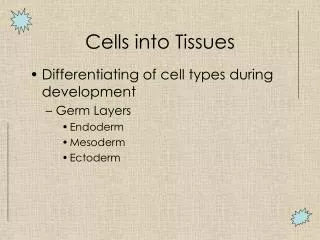
3-layered Flat Embryo Ectoderm (1) Mesoderm (3) Endoderm (2) Fertilized egg or Zygote (Single cell) 3-layers of cells All Tissues & Organs of Human body
Human Genome Nucleus Cytoplasm Cell membrane Cell Each cell has 46 chromosomes Form 23 homologous pairs Each parent contributes = 23 Autosomes = 44 Sex chromosomes = 2 (Male = XY, Female = XX) Chromosomes (2N = 46) Each autosome of a homologous pair look alike But each sex chromosome do not look alike
Cell Cycle Paclitaxel Vincristine Vinblastine Colchicine Nondividing cells (Fixed postmitotics) M G0 Resting cells (Reverting postmitotics) G1 G2 Bleomycin Etoposide S Methotrexate
Mitosis & Meiosis A Homologous Pair (2 Chromosomes) 1 2 46 1 pair 23 pair 1st Division 2nd Division Daughter Somatic Cells (2) Gametes (4)
Meiosis Takes place only in testes and ovaries Is a reductional division Main purpose is to reduce the number of chromosomes from 2N to 1N in sperms & eggs (Chromosomes of each homologous pair will separate from each other) Homologous pair = 1 chromosome from each parent (at fertilization) 2N = 46 chromosomes (2 sets) 1N = 23 chromosomes (1set) So Sperm = 1N chromosomes (23) Egg = 1N chromosomes (23) Fertilization restores chromosome number again to 2N = 46 chromosomes (2 sets)
Human Body Cells Tissues Organs Human body Cell Tissue
Cell Organelles • Nucleus Chomatin, Transcription • Rough ER Protein synthesis & Segregation • Smooth ER Fat & Steroid synthesis & Detoxification • Golgi Complex Concentrating, Modifying & Packaging of secretory products • Lysosomes Intracellular digestion • Peroxisomes Contain oxidative enzymes; Use catalase to degrade H2O2 = H2O + O2 • Mitochondria Oxydative phosphorylation & ATP production • Cell Membrane Lipid bilayer layer with intramembranous proteins • Cell cytoskeleton Actin filaments, Microtubules, intermediate filaments
Cell Organelles Mitochondria Lysozome Golgi Nucleus Rough ER
Cells, Tissues & Various Topics Of Research • Subcellular localisation & trafficking of molecules and oganelles • Cell-cell and cell-extracellular matrix interactions • Cell cytoskeleton and receptor dynamics and functions • Cell and tissue differentiation and remodelling • Genetically engineered cells and tissues • Three-dimensional reconstructions, particularly of expression patterns over time • Cell cycle and cell lineage analysis involving gene expression profiles • Apoptosis • Gene expression analysis from histological preparations • Functional genomics & proteomics • Techniques used in molecular histology
Epithelial Tissue Outer layer of skin Inner lining of trachea Inner lining of ducts of sweat glands
General Features • Diversity • Metaplasia • Lining and Covering • Basal Lamina • Renewal • Avascularity • Cell Packing • Derivation
Classifying Principles 1. Number of cell layers: 1. Simple epithelia 2. Stratified epithelia 3. Pseudostratified epithelia 2. Shape of the surface cells: 1. Squamous cells 2. Cuboidal cells 3. Columnar cells 3. Luminal surface modifications: 1. Microvilli (Brush border) 2. Cilia 3. Stereocilia
Specific Epithelial Types • Simple squamous epithelium: • Simple cuboidal epithelium: • Simple columnar epithelium: • Pseudostratified epithelium: • Stratified Squamous epithelium: a) Keratinized b) Nonkeratinized • Stratified cuboidal epithelium: • Stratified columnar epithelium: • Transitional epithelium:
Types of Epithelia Simple squamous Stratified squamous Transitional Simple cuboidal stratified cuboidal Full Empty Bladder Simple columnar Pseudostratified
Kidney (Epithelium) Simple squamous Simple cuboidal Kidney Tubules
Small Intestine (Simple Columnar) Absorptive cells Nucleus Brush border Lamina propria Lumen of gut
Esophagus (Stratified Squamous) Epithelium Lamina propria
Skin (Stratified Squamous) Epidermis (Epithelium) Dermis (Connective tissue)
Trachea (Pseudostratified Epithelium) Epithelium Cilia Ciliated cells Goblet cells Basal lamina Lamina propria
Ureter (Transitional Epithelium) Epithelium Lumen Basal lamina Lamina propria
Basal Lamina • Next to epithelia an acellular sheet like structure is the Basal Lamina. • Component Layers & Constinuent Macromolecules: A. Component Layers Lamina lucida Lamina densa B. Constituent Macromolecules Lamina lucida (Laminin that binds to cell surface integrins, collagen IV) Lamina densa (Type IV Collagen) Basement Membrane: Basal lamina accompanied by reticular lamina (Type III Collagen) is called the basement membrane. Functions: Forms sieve-like selective barrier between the epithelia & connective tissue. Aids in cell organization, cell adhesion & maintainence of cell shape. Has a role in maintaining specific cell function. Helps guide migrations of cells during development and regeneration of injured tissue
Polarity & Specialization of Epithelial Cells • Specialization of the Apical Surface: 1. Microvilli (Enterocytes & Proximal convoluted tubule cells)) 2. Cilia (Trachea, Bronchus etc.) 3. Stereocilia (Epididymis) 4. Flagella • Specialization of the Lateral Surfaces: 1. Zonula occludens (Tight junctions) 2. Zonula adherens (Intermediate junctions) 3. Macula adherens (Desmosomes) 4. Gap junction (Nexus) • Specialization of the Basal Surface: 1. Basal lamina 2. Hemidesmosome 3. Sodium-potassium ATPase D. Intracellular Polarity:
Cell Junctions Microvilli Zonula occludens Zonula adherens Terminal web Macula adherens Gap junction Nucleus Hemidesmosome
Mucous Membranes • Components of Mucous Membrane: 1. Epithelium 2. Basement membrane 3. Lamina propria
Mucous Membrane Epithelium Basal lamina Lamina propria
Serous Membranes • Components of Serous Membrane: 1. Epithelium called mesothelium 2. Basement membrane 3. Submesothelial connective tissue layer
Functions of Epithelia 1. Protection from: Mechanical trauma Dehydration Pathogens • Secretion of: Hormones, milk, sweat etc. Enzymes, HCl, glycoproteins, Mucous & serous products • Lubrication of: Contents of GI tract Fetus in birth canal Joints 4. Filtration of wastes: (Urine) • Absorption of food: (Aminoacids, Glucose, Fatty acids) • Neuroepithelium: (Taste, Smell, Hearing) • Reproduction: (Germ cells)
Major Types of Epithelial Cells • Epithelial Cells Specialized for Transport: 1. Ion-transporting cells (Kidney tubules, Gall bladder etc.) 2. Cells that transport by pinocytosis (Endothelial cells of blood capillaries • Absorption: (Enterocytes, Proximal convoluted tubule cells) • Secretion: 1. Protein-secreting cells (Acinar cells of pancreas, Hepatocytes) 2. Polypeptide-secreting cells (APUD cells) 3. Mucous cells (Goblet cells) 4. Serous cells (Acinar cells of pancreas & secretory cells of parotid salivary glands. 5. Steroid-secreting cells (Adrenal cortex, Leydig cells etc.) D. Contractile Epithelial Cells: (Myoepithelial cells of glands)
GLANDS • Exocrine & Endocrine Glands: • Classification of Exocrine Glands: 1. By structure: a) Number of cells b) Duct system c) Secretory portion 2. By secretory product a) Mucous secretion b) Serous secretion c) Seromucous secretion 3. By mode of secretion a) Merocrine b) Apocrine c) Holocrine
Unicellular Multicellular Simple tubular Coiled tubular Branched Simple branched Simple acinar Compound tubular Compound tubulo-alveolar
Salivary Glands Mucous acini Serous acini
Mode of Secretion Active transport Merocrine Apocrine Holocrine Endocrine
Connective Tissue Fat Fat cells Tanden Fibroblasts Bone Osteocytes
Connective Tissue • Is one of the 4 basic tissues of the body. • Structurally it is made up of cells and large amount of intercellular space containing extracellular matrix. • Matrix is the dominating component of this tissue. • It forms framework, connecting, supporting and packing tissue of the body. • It also plays a dynamic role in the development, growth and homeostasis of other tissue types.
Connective Tissue Loose connective tissue Dense connective tissue Fibroblasts Extracellular matrix Epithelial tissue Mammary Glands
Composition • Cells • Extracellular matrix
Types of Cells in Loose Connective Tissue • Residents: Fibroblasts Macrophages Reticular cells Mesenchymal cells • Visitants: Mast cells Plasma cells Leukocytes Fat cells Melanocytes
Loose Connective Tissue Elastic fibers Capillary Neutrophil Plasma cell Fibroblast Collagen fibers Macrophage Adipocyte Mast cell Lymphocyte
Fibroblast (Ultrastructure) Nucleus Rough ER Collagen Extracellular matrix
Collagen Producing Cells • Fibroblast-More than one type of collagen • Chondroblast- Type II collagen • Osteoblast-Type I • Reticular cell- Type III • Smooth muscle-Type I & III
Extracellular Matrix • Extracellular matrix (Fibers & Ground substance) is synthesized and secreted mainly by the fibroblasts & the fibers are assembled in the extracellular space. • Fibers Prime function is support & plays strengthing role in • Ground substance Functions are 1. Acts as a molecular sieve & stops the spread of noxious substances 2. Plays very important role in cellular nutrition & waste removal 3. Plays a vital role in aging. Its amount diminishes with age and wrinkles start appearing.
Fiberous Components Connective tissue fibers are long, slender protein polymers that are present in variable proportions in different types of connective tissue. In many cases the predominant fiber type is responsible for conferring specific properties on the tissue. • Collagen Fibers: • Elastic Fibers: • Reticular Fibers:
Collagen Fibers Collagen Fibers: Most abundant protein in the body. Synthesis & assembly: Collagen types- Type I- most abundant & occurs in loose and dense connective tissue & bone. Type II- occurs in cartilage. Type III- occurs in hematopoitic tissues. Type IV- occurs in basal laminae & does not form fibers or fibrils. Type V- in placental basement membranes & blood vessels. Type X- around hypertrophic, degenerating chondrocytes of the growth plate where bone formation is to occur.
Synthesis of Collagen Collagen’s main amino acids Glycine (34%) Proline (12%) Hydroxyproline (10%) Fibroblast Procollagen (Triple-helical units) Procollagen peptidase Tropocollagen Collagen fibril Collagen fiber Intracellular Extracellular
Ground Substance Proteoglycans: They are made up of a core protein to which glycosoaminoglycans (GAGs) are attached. GAGs are polysacharides that contain aminosugars. GAGs-Chondroitin sulphate, Dermatan sulphate, Keratan sulphate & Heparin sulphate. Hyaluronic acid is a GAG but do not form proteoglycans. Matrix viscosity and rigidity are determined by the amount and types of GAGs, their association with the core protein to form proteoglycans, GAG-fiber association, and GAG-GAG associations. Glycoproteins: Fibronectin-mediates the attachment of cells to the extracellular matrix. Laminin-a component of basal laminae that mediates the attachment of epithelial cells. Tissue fluids: Salts:
Connective Tissue Types A. Connective Tissue Proper: 1. Loose connective tissue 2. Dense connective tissue a) Dense regular connective tissue b) Dense irregular connective tissue • Reticular connective tissue: • Elastic connective tissue: • Mucous connective tissue:
Connective Tissue Proper A. Connective Tissue Proper: 1. Loose connective tissue (lamina propria) 2. Dense connective tissue a) Dense regular connective tissue (Tendon, ligament) b) Dense irregular connective tissue (Dermis, organ capsule) Loose CT Dense CT