
The Nervous System
The Nervous System. I. Functions of the Nervous System. A. Nervous system is our processing system, and the system that keeps us in contact with the outside world . Intro Animation B. Roles of the nervous system: 1. Coordination of movement 2. Response to environmental stimuli

The Nervous System
E N D
Presentation Transcript
I. Functions of the Nervous System A. Nervous system is our processing system, and the system that keeps us in contact with the outside world. Intro Animation B. Roles of the nervous system: 1. Coordination of movement 2. Response to environmental stimuli 3. Intelligence 4. Self-awareness (consciousness) 5. Thoughts 6. Emotions
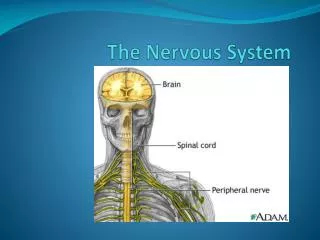
II. Composition of the Nervous System A. Made up of nerve cells called NEURONS which are specialized to carry nerve impulses
Neurons: Structure and Function I. Parts of a Neuron A. Dendrite: conducts message towards the cell body. Animation
B. Axon: conducts message away from the cell body.
C. Synapse: a junction between neurons; where message jumps from one neuron to another.
F. Node of Ranvier: gaps in the myelin sheath, they speed up transmission of nerve impulses.
G. Cell body: conducts the normal metabolic functions of the cell.
Types of Neurons: Structure and Function I. Motor Neuron (Efferent Neuron) A. Connected to an effector(muscle fibre or gland). B. Appearance 1. Short dendrite 2. Long axon 3. Cell body positioned inside the central nervous system (CNS).
C. Transmitted impulses cause effector to react (eg. muscle to contract). D. Message is traveling away from the brain
II. Sensory Neuron (Afferent neuron) A. Starts with a sensory receptor (pressure, heat, light, etc.). B. Appearance 1. Long dendrite 2. Short axon 3. Cell body is outside CNS in ganglia
C. Transmitted impulses send information about the environment to the brain. D. Message travels towardsCNS. Animation
III.Interneuron (Association neuron) A. Smaller than other two types of neurons. B. Located entirely within CNS. C. Usually, short dendrites. D. Axons can be long or short. E. Conveys messages between system parts in CNS.
Transmission of Nerve Impulses • I. Nerve Conduction A. Occurs due to an electrochemical change that moves in one direction along the length of a nerve fiber. B. It is electrochemical because it involves changes in voltage as well as in the concentrations of certain ions.
Three Phases of Nerve Impulse • A. Resting Potential • 1. Potential difference across the membrane of the axon when it is not conducting an impulse is -60 mV. • 2. This negative polarity is caused by the presence of large organic negative ions which are too large to cross the membrane.
3. During the resting potential, Na+ ions are more concentrated on the outside of the membrane than the inside. 4. K+ ions are more concentrated on the inside of the axon. 5. This uneven distribution of K and Na ions is maintained by active transport across Na+/K+ pumps which operate whenever the neuron is not conducting an impulse. 6. Must work all the time, because themembrane is partially permeable to these ions, and they tend to diffuse toward regions of lower concentration
B. Action potential 1. If a nerve is stimulated by electric shock, pH change, mechanical stimulation, a nerve impulse is generated, and a change in potential can be seen. 2. This nerve impulse is called the ACTION POTENTIAL. 3. Broken into an upswing and downswing. a. During the upswing (-60 mV to +40 mV), membrane becomes permeable to Na+ ions. i. Na+ ions move from outside to inside of axon due to opening of the sodium gates in the membrane. ii. Depolarization occurs because the inside of the axon becomes positive.
b. In the downswing (+40 mV to -60 mV), membrane becomes permeable to K+ ions. i. K+ ions moves from outside to inside of axon due to the opening of the potassium gates in the membrane. ii. Repolarization occurs because the inside of axon becomes negative again.
C. Recovery Phase 1. Occurs between transmissions, K+ ions are returned to inside of axon, Na+ to the outside. 2. This is done actively by the Na+/K+ pumps. 3. Neuron is now ready to send another impulse! 4. Multiple impulses can pass down a nerve in succession. 5. Only small amounts of the ions move, and the resting potential is quickly restored.
III. Summary of Nerve Impulse Transmission A. The 3 phases of nerve impulse. 1. Resting potential 2. Action potential – depolarization event (Na+ gates opening, Na+ pouring in) 3. Action potential – repolarization event (K + gates opening, K + pouring out) AnimationAnimation 2 4. Resting potential – neuron is ready to conduct again.
B. Notes about transmission 1. Impulse transmission is one way only. a. As depolarization wave passes along, it stimulates the sodium gates to open at the very next point. b. Gates that have just opened and closed cannot be restimulated for a very brief period of time. c. This is called a REFRACTORY or Recovery Period. d. Makes it impossible for the impulse to travel “upstream”.
direction+- - - - - - - - - - - - - - - - r r +- - - - - - - - - - - - - - - - r r +- - - - - - - - - - - - - - - - r r +- - - - - - etc.
2. Impulses travel from receptor (or dendrite) down the axon. 3. The impulse, once triggered, is the same each time, and in every neuron (-60 to +40 mV). 4. A neuron is either transmitting, or it is not. a. Often called the “all-or-none” response. b. A stimulus must meet or exceed the neuron’s THRESHOLD for triggering depolarization, or there is no impulse sent … but neurons do not distinguish between “meet” or “exceed.” Animation
Myelin Sheath I. Description A. Composed of lipids. B. Composed of tightly packed spirals of the cell membrane of Schwann cells. C. This sheath gives nerves their characteristic shiny white appearance.
II. Function A. Acts as insulation B. Speeds up impulse transmission 1. The speed of transmission is ~200 m/s in myelinated fibers, but only 0.5 m/s in non-myelinated fibers. 2. The reason is that the nerve impulse "jumps" from node to node in myelinated fibers (Saltatory conduction.)
3. In non-myelinated fiber, the nerve impulse must depolarize and repolarize each point along the nerve fiber. Animation
The Synapse I.Why are Synapses Important? A. A SYNPASE is specialized region at the ends of axons called synapses allow one nerve cell to communicate and transmits an impulse across to another cell.
A.Synapse: the region between end of an axon and the cell body or dendrite to which it is attached. B. Synaptic Endings: swollen terminal knobs on the ends of axon terminal branches. C. Presynaptic Membrane: the membrane of the axon synaptic ending. D. Postsynaptic Membrane: the membrane of the next neuron just beyond the axon's synaptic membrane.
E. Synaptic Cleft: the space between the presynaptic and the postsynaptic membranes F. Neurotransmitter Substances (neurotransmitters): chemicals that transmit the nerve impulses across a synaptic cleft. 1. Some important neurotransmitters a. Acetylcholine (Ach) b. Noradrenalin (NA) c. Serotonin d. Adrenalin (epinephrine) G. Synaptic Vesicles: contain the neurotransmitters. Contained near surface of synaptic endings.
Synaptic Transmissions A. An action potential reaches the axon bulb B. Ca2+ gates in bulb membrane open. C. Entry of Ca2+ bulb stimulates the synaptic vesicles to move to the presynaptic membrane. D. They fuse with the membrane, emptying the neurotransmitter into the synaptic cleft by exocytosis
E. Neurotransmitters diffuses across the cleft to the receptors on the postsynaptic membrane of the next neuron’s dendrite. F. The action of binding the neurotransmitters initiates or suppresses an action potential in the postsynaptic neuron. AnimationAnim 2
III. Notes about Impulses Across the synapse A. Impulses can only go one way across the gap because only the axon has the vesicles and the dendrite only has the receptors. B. Different nerve cells use different chemicals as neurotransmitters. C. Most neurotransmitters are EXCITATORY –their binding opens Na+ channels in the membrane, and creates or encourages action potentials.
D. Some neurotransmitters are INHIBITORY – their binding stops action potentials. E. Exact action depends more on the receptor than on the neurotransmitter. a. E.g. Serotonin can be excitatory or inhibitory in different circuits.
F. A single neuron may receive information from thousands of neighbouring neuron through thousands of synapses. 1. Some of the messages are excitatory (i.e. they tell the neuron to “fire”) while others may be inhibitory (i.e. they tell the neuron not to fire). 2. Whether or not a neuron “fires” off an action potential at any particular instant depends on its ability to integrate these multiple positive and negative inputs. 3. This allows neurons to be fine-tuned to the environment
Neurotransmitters • Neurotransmitters take nerve impulses across synapses. • Neurotransmitters are small molecules. • They can be single amino acids, short chains of amino acids, or derivatives of protein. • Proper brain and nervous system function depends on the proper balance of excitatory and inhibitory synaptic transmitters. • Excitatory transmitters include 1. Acetylcholine (ACh) 2. Adrenalin (epinephrine) 3. Noradrenalin (norepinephrine) 4. Serotonin (derived from the amino acid tryptophan) 5. Dopamine
F. Inhibitory transmitters include 1. GABA (gamma aminobutyric acid - a type of amino acid) 2. Glycine (an amino acid) 3. Serotonin can also act as an inhibitory neurotransmitter. • Neurotransmitters include endorphins and enkephalins (a 5 amino-acid chain that functions as a natural pain reliever in brain). • Opium and heroin mimic the action of natural endorphins and enkephalins. Animation
Neurotransmitters Breakdown A. Inactivating neurotransmitters are important to clear the synaptic gap for the next signal from the presynaptic neuron. B. Methods to remove neurotransmitters: 1. Enzymes exist in the synaptic cleft to break apart the neurotransmitter (e.g. acetyolcholinesterase) 2. Reabsorption of neurotransmitters by axon bulb for breakdown or repackaging/reuse.
Reflex Arc I. Reflex A. Reflexes are automatic, involuntary responses to changes occurring inside or outside the body. B. Can involve the brain (e.g. blinking)or not involve brain (e.g. withdraw hand from hot stove). C. The reflex arc is the main functional unit of the nervous system and allows us to react to internal and external stimuli Animation
II. Parts of a Simple Reflex Arc II. Parts of a Simple Reflex Arc A. Receptor (e.g. in skin) - generates a nerve impulse. B. Sensory Neuron - takes message to CNS. Impulses move along dendrite, proceed to cell body (in dorsal root ganglia) and then go from cell body to axon in gray matter of cord.
Interneuron- passes message to motor neuron. Motor neuron - takes message away from CNS to axon of spinal nerve. Effector- receives nerve impulses and reacts: glands secrete and muscles contract.
III. How the simple reflex arc works A. Receptor receives info from the environment, and initiates a nerve impulse. B. An interneuron in the spinal cord bypassesthe brain and sends impulse directly to a motor neuron. C. Motor neuron transmits impulse to an Effector. (e.g. muscle fibre) D. Protective action initiated. E. Interneuron does “notify” the brain of the stimulus but by the time the brain can respond, the reflexive action has already occurred!


