Download
1 / 9
90 likes | 274 Vues
TOPEM TOF- PET MRI for prostate cancer diagnosis and follow up Sezioni partecipanti: Roma1, Gr. Coll. ISS, Genova, Bari, LNS

E N D
TOPEM TOF- PET MRI for prostate cancer diagnosis and follow up Sezioni partecipanti: Roma1, Gr. Coll. ISS, Genova, Bari, LNS Il cancro della prostata e’ il piu’ diffuso e la seconda causa di morte per l’uomo. Non esistono tecniche affidabili per la diagnosi la caratterizzazione ed il follow up. Le tecniche in uso (PSA, DRE. Ecografia, MRI, CT) non sono sufficientemente sensibili e/o specifiche. Si fa sempreanche la biopsia, tecnica invasiva ed eseguita “al buio” (unico caso nel campo della diagnosi del cancro). E’ di fondamentale importanza la precisa localizzazione e stadiazione della neoplasia e la valutazione della sua aggressività biologica. E’ necessario combinare tecniche morfologiche (MRI, trattandosi di tessuto molle) con tecniche funzionali (PET) ed aumentare in modo considerevole le prestazioni dei rivelatori PET in termini di risoluzione spaziale ed efficienza. Scopo dell’esperimento e’ il progetto e la costruzione di un “sistema diagnostico” costituito da una sonda endorettale PET, compatibile con MRI, che puo’ essere utilizzata in coincidenza con una PET standard esterna e/o con uno o piu’ moduli PET dedicati. Le performances del sistema sono dominate da quelle del probe endorettale ad alta risoluzione ed alta efficienza vicinissimo alle lesioni. Si possono ottenere quindi risoluzioni spaziali di 1 mm o poco piu’ ed elevata efficienza (PET standard 4 – 6 mm). La tecnica TOF consente di aumentare l’SNR (NECR) (quindi la rivelabilita’ della lesione), tanto piu’ quanto migliore e’ la risoluzione in timing (≤ 300 ps FWHM). La multimodalita’ in contemporanea con MRI potenzia enormemente le capacita’ diagnostiche del sistema). Si deve costruire un sistema finalizzato cioe’ nella giusta scala per rendere possibile il trasferimento tecnologico.
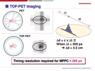
TOPEM (PET TOF probe, compatible with MRI and MRS for diagnosis and follow up of prostate cancer) F. Garibaldi - c.d. s. Roma1, 09-07-2010 Prostate cancer is the most common cancer and thesecond leading causeof cancer death SENSITIVITY 83%SPECIFICITY 17% PSA it is the only case for cacer diagnosis made from tissue obtained on a blind biopsy !! In practice there is nothing reliable for diagnosis and follow up • - detectors far away from prostate • -poor spatial resolution (6 – 12 mm) • poor photon detection efficiency (<1%) • activity ouside the organ • -> poor contrast resolution Multimodality approach is needed drawback of the standard PET The solution PET – TOF MRI endorectal probe, coupled to external detector(s) and/or standard PET TOF (< 300 ps FWHM ! ) would solve the problem huge background from the bladder Could we reduce or eliminate it?
Why TOF ? Improvements in clicnical images (600 ps) - TOF provides huge performance increase! Can localize the source along line of flight. With 300 ps FWHM, 4.5 cm (just the dimension of the prostate) (in a certain sense one doesn’t “see” the bladder) - Fast convergence (reduced scan time) First results from Catania (LNS) We did better !
Limited space for the PET detector • PET detector must not use magnetic materials • -> Could distort MR imagge • PET detector must not emit in MR frequency • Could produce MR image artifacts • MR-compatible PET shielding materials • Could distort MR image • MR gradient field-eddy currents • Could produce noise in detector • Could heat detector • MR RF transmit • Could produce false PET events • MR materials • Will produce more gamma attenuation - CITRATE that is present in the normal prostate -CREATINAthat may increase in the phlogosis and all the proliferative processes -COLINEspecific for a neoplastic transformation Effetti della PET su MRI e viceversa I problemi dell’impatto relativo PET-MRI sono risolti nostri test primi Settembre a Tor Vergata B. Pichler et al. “Simultaneous PET-MRI, a new approach “ Nature Medicine Vol 14, N 4, April 2008 In conclusion, our results confirm that simultaneous PET and high-field-strength MR imaging with LSO-APD–based PET detectors is feasible without sacrifici the quality of images obtained with either system. The next step will be to focus on the evaluation of the full-ring PET insert. While this work was concentrated on small-animal PET/MR imaging, our results can be transferred to a clinical system, where MR imaging is usually performed at a much lower magnetic field strength. The combination of PET and MR imaging can open new opportunities in preclinical research and clinical diagnosis. When nuclear MR spectroscopy is used with MR imaging and PET, trimodal imaging might be possible and thereby add even more information in biomedical research studies, a new way for a new perspectives in molecular imaging
Elettronica • Discreta (NINO + HTDC) • ASIC – 1 front end • - ASIC - 2 (con TDC) • Elettronica integrata Layout
Milestones concordate per il 2011 • 15 Luglio: test del front end dell'ASIC e sua integrazione nella scheda ibrida- progetto e costruzione della prima scheda ibrida "finale” • OK • 31 Dicembre: costruzione del probe (prototipo "zero") e dei rivelatore in coincidenza. Studio della risoluzione energetica, risoluzione temporale ed eventualmente della DOI con cristalli a differente ruvidità superficiale • Si e’ costruito il minirivelatore che e’ attualmente in fase di test a Genova con elettronica ibrida
Milestones proposte per il 2012 - 15 Luglio 2012 1. progetto e costruzione dell'ASIC che integra il TDC a 32 canali 2. progetto e costruzione delle schede della nuova elettronica miniaturizzata 3. caratterizzazione di cristalli (LSO, LYSO, LSF) ,misure DOI + TOF con PMT veloci e SIPM 4. misure di PDE di SiPM diversi - 31 Dicembre 2012 5. prime misure di timing con elettronica discreta (NINO + HTDC) 6. integrazione dell'ASIC nella nuova elettronica, test del sistema, misure preliminari di timing 7. costruzione del contenitore del probe, del sistema di raffreddamento e monitoring della temperatura Nessuna difficolta’ per le milestones 2, 3, 4, 5 (per 4 e 5 si e’ gia’ fatto molto). Le milestone 1 un finanziamento pari a quello richiesto entita’. Per la milestone 7, il sistema di raffreddamento e monitoring della temperatura non e’ stato finanziato. La collaborazione e’ orientata a chiedere un prolungamento dell’esperimento al 2013. Cio’ consentirebbe di costruire un sistema finalizzato cioe’ nella giusta scala per rendere possibile il trasferimento tecnologico.