Hyperparathyroidism
Hyperparathyroidism. PTH/Calcium Homeostasis.

Hyperparathyroidism
E N D
Presentation Transcript
PTH/Calcium Homeostasis Low circulating serum calcium concentrations stimulate the parathyroid glands to secrete PTH, which mobilizes calcium from bones by osteoclastic stimulation. PTH also stimulates the kidneys to reabsorb calcium and to convert 25-hydroxyvitamin D3 (produced in the liver) to the active form, 1,25-dihydroxyvitamin D3, which stimulates GI calcium absorption. High serum calcium concentrations have a negative feedback effect on PTH secretion.
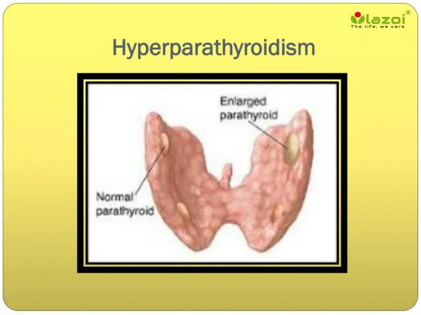
Parathyroid glands-Surgical anatomyEndocrine glands. Usually2 on each side i.e 4 in No(84%, >4 in 13%, <4 in3%..2 sup .glands: parathyroid IV, derived from 4th pharyngeal pouch.constant in position ,behind RLN 2inf PG.variable in position, derived from 3rd pharyngeal pouch, hence called parathyroid III. Usually lie ant. toRLN. Each gland: 5 to 7 mm(6X2X2 mm).Wt 40-50mg.yellowish-Brown firm gland which sinks in water unlike fat (Float) Blood supply:Mostly from inf thyroid art.-entering a hilumlikestructure,afeaturethat differentiates it from fat Venous drainage- ipsilat.toSup,middle,inf TV
I.Hyperparatyhyroidism-Primary hyperparathyroidismTertiary HPT II. Malignancy-related-Solid tumor with metastases (breast)-Solid tumor with humoral mediation of hypercalcemia (lung, kidney)-Hematologic malignancies (multiple myeloma, lymphoma, leukemia) III. Endocrine diseases:Hyperthyroidism.Addisonian crisis.pheochromocytoma IV- Granulomatous diseases:Sarcoidosis.T.B. IV. Iatrogenic:Excessive intake of Vit D or calcium-Rx with lithium-Thiazide diuretics V. Associated with renal failure-Severe secondary hyperparathyroidism-Aluminum intoxication VI-Familial hypocalcuric hypercalcemia-Milk-alkali syndrome Hypercalcemia **Primary hyperparathyroidism and cancer account for 90% of cases of hypercalcemia
PHPT Incidence :0.1-0.3%. 1 case per 1000 men and 2-3 cases per 1000 women.25/100000 population Incidence increases above age 40 Most patients with sporadic PHPTarepostmenopausal women with an average age of 55 years Etiology: a solitary parathyroid adenoma(83%) Multiple adenomas (6%) Hyperplasia 10% Carcinoma 1% Primary Hyperparathyroidism
Primary HPT: Clinical Features • Symptomatic: • Classical pentad of symptoms(Kid.stones, • painful bones,abdominalgroans,psychic moans,& fatigue overtones) • Osteitisfibrosacystica • Nephrolithiasis • Pathologic fractures • Neuromuscular disease • Life-threatening hypercalcemia • DU.pancreatitis ?Asymptomatic:Hypercalmic • Fatigue, muscle weakness & ache • Depression • Polydipsia.Polyuria • Anorexia,dyspepsia wt loss.Constipation • SOB.HT
Investigations Intact PTH and chemistry panel • PTH elevated (normally 0.15-1ng/ml)despite elevated serum calcium.(fasting + no cuff on arm on sampling) • Serum phosphate decreases. Alkaline phosphatase elevated • Look at the serum creatinine to evaluate for CRI/CRF • Rule out lithium or thiazide use • 24-hour urine calcium excretion • Used to rule out familial hypocalciuric hypercalcemia • Values below 100mg/24 hours or a calcium creatinine clearance ratio of <0.01 are suggestive of FHH • XR-Skull; salt pepper appearance.;Subperiosteal erosion of radial side of middle phalanx.Osteitis fibrosa cystica. • Consider KUB, IVP or CT to evaluate for kidney stones • Ionized calcium versus serum calcium—the debate rages on…. • CORRECTED SERUM CALCIUM • Serum calcium (mg/dL)+(0.8X[4-albumin (g/dL)])
Surgical Candidacy • Symptomatic primary HPT • Serum calcium greater than 1mg/dL above the upper limit of the reference range(>11mg%) • 24 hour urine calcium greater than 400 mg • Creatinine clearance reduced by more than 30% compared with age-matched subjects • Marked reduced Bone density • Age under 50 • -Urinary calculi • Neuromascular presentation
general Considerations in Mx PRICIPLES OF Mx; I-Clinically_ 1-Do symptoms due to hypercalcemia Or no?t.Duration?. 2- symptoms related to malignant disease or not Recent cough,wheeze,or hemoptysis—consider Bronchogenic Ca. Hematuria may suggest:hypernephroma, bladder Ca. or Renal stones 3-Conditions associated with HPT?.-Renal colic, PU,Pancreatitis,,HT,Gout. 4-Possible excess use of milk products,antacids,baking soda,or vitamins II-Biochemically-Lab High S. Calcium+S.Phosphate-----Suggest HPT High S. Calcium + Normal S. phosphate( in50% of HPT,& may also in patients with Vit D intoxication,sarcoidosis,malig dis without metastasis& hyperthyroidism S.Chloride: S. Phosphate ratio >33 suggestsHPT Measure S. Parathyroid hormone- low or normal in all causes of hypercalcemia other than Prim HPT or Benign familial hypocalcuric hypercalcemia(BFhH).Chronic hypercalcemia+mildly elevated PTH level+ low urinary Calcium+family Hy of hypercalcemia esp.in children III-Localization-Usually required in persistent or recurrent HPT US-localize the tumors in 75%. &SESTAMBI scan in 85%..These studies localize in 35% of Hyperplasia Experienced surgeon can find tumors in 95 % without these tests in 1st op.
IV-post-op Bone hunger- In 24-48h-hypocalcemia’ Mild oral Calcium+ active vit D3(one alpha-leo Vit D3-0.5μg bid Severe – I.V Calcium gluconate(10ml) slowly over 10mint or infusion
Pre-Operative Imaging-Localization • High-resolution ultrasound • Sensitivity 65-85% for adenoma; 30-90% for enlarged gland • Results suboptimal in pts with multinodular thyroid disease, pts with short thick neck, ectopic glands (15-20%) • May be useful in detecting sestamibi scan negative adenomas • CT with contrast/thin section • Sensitivity of 46-87% • Good for ectopic glands in the chest • MRI • Sensitivity of 65-80% • Good for ectopic glands • Sestamibi • 85-95% accurate in localizing adenoma in primary HPT • Sestamibi-SPECT(single photone emission CT) • Sensitivity 60% for enlarged gland and 98% for solitary adenomas
Scintigraphy Images Traditional Sestamibi Sestamibi-SPECT
Medical Management • Asymptomatic patients may elect to be closely followed and managed medically • A recent study of pts with asymptomatic primary HPT showed that the majority of pts followed for ten years did not demonstrate an increase in serum calcium or PTH levels—25% of patients had progressive disease including worsening hypercalcemia, hypercalciuria and reduction in bone mass—younger patients more likely to have progression of disease • Patients opting not to have surgery should have a serum calcium level drawn every 6 months and should have annual bone densiometry at all three sites
Medical Management Primary HPT • Estrogen • Dose required is high • SERMs • Reduction in serum calcium and markers of bone turnover after 4 weeks • Bisphosphonates • Studies have shown increase in lumbar spine and femoral neck mineral density • Calcium/Vitamin D • Calcimimetic agents (Cinacalcet) • Under investigation for primary HPT
Familial Syndromes • MEN I • MEN IIA • Familial Hypocalciuric Hypercalcemia • Hyperparathyroidism-jaw tumor syndrome • Fibro-osseous jaw tumors • Renal cysts • Solid renal tumors • Familial isolated hyperparathyroidism
MEN I • MEN I • 1 in 30,000 persons • Features: • Hyperparathyroidism (95%) • Most common and earliest endocrine manifestation • Gastrinoma (45%) • Pituitary tumor (25%) • Facial angiofibroma (85%) • Collagenoma (70%) • HPT in MEN I • Early onset • Multiple glands affected • Post-op hypoparathyroidism more common (more extensive surgery) • Successful subtotal parathyroidectomy followed by recurrent HPT in 10 years in 50% of cases
STIGMATA OF MEN I Lipomas Collagenomas Angiofibromas
MEN IIA (Sipple’s Syndrome) • Features: • MTC(95%) • Pheochromocytoma(50%) • HPT(20%) • RET mutation (98%) • 1 in 30,000-50,000 people • Usually single adenoma but may have multi-gland hyperplasia
Familial Hypocalciuric Hypercalcemia This benign condition can be easily mistaken for mild hyperparathyroidism. It is an autosomal dominant inherited disorder characterized by hypocalciuria (usually < 50 mg/24 h), variable hypermagnesemia, and normal or minimally elevated levels of PTH. These patients do not normalize their hypercalcemia after subtotal parathyroid removal and should not be subjected to surgery. The condition has an excellent prognosis and is easily diagnosed with family history and urinary calcium clearance determination.
Secondary Hyperparathyroidism • Decreased GFR leads to reduced inorganic phosphate excretion and consequent phosphate retention • Retained phosphate has a direct stimulatory effect on PTH synthesis and on cellular mass of the parathyroid glands • Retained phosphate also causes excessive production and secretion of PTH through lowering of ionized Ca2+ and by suppression of calcitriol production • Reduced calcitriol production results both from decreased synthesis due to reduced kidney mass and from hyperphosphatemia. • Low calcitriol levels, in turn, lead to hyperparathyroidism via both direct and indirect mechanisms. Calcitriol is known to have a direct suppressive effect on PTH transcription and therefore reduced calcitriol in CRD causes elevated levels of PTH • Reduced calcitriol leads to impaired Ca2+ absorption from the GI tract, thereby leading to hypocalcemia, which then increases PTH secretion and production.
Secondary HPT • Clinical presentation • Usually asymptomatic • Diagnosis • Elevated PTH in the setting of low or normal serum calcium is diagnostic • If phosphorous is elevated, cause is renal • If phosphorous is low, other causes of vit D deficiency should be sought • Prevention • Vit D replacement • Phosphorus binders [Sevelamer] • Treatment • Medical • Calcimimetic agents • Surgical • Considered in cases of refractory severe hypercalcemia, severe bone disease, severe pruritis, calciphylaxis, severe myopathy
Tertiary Hyperparathyroidism Tertiary hyperparathyroidism develops in patients with long-standing secondary hyperparathyroidism, which stimulates the growth of an autonomous adenoma. A clue to the diagnosis of tertiary hyperparathyroidism is intractable hypercalcemia and/or an inability to control osteomalacia despite vitamin D therapy. Surgical Referral - calcium- phosphate product > 70 - severe bone disease and pain -intractable pruritus - extensive soft tissue calcification with tumoral calcinosis -calciphylaxis
Lab Abnormalities • Primary HPT • Increased serum calcium • Phosphorus in low normal range • Urinary calcium elevated • Secondary HPT (renal etiology) • Low or normal serum calcium • High phosphorus • Tertiary HPT (renal etiology) • High calcium and phosphorus
Hypercalcemic crisisHypercalcemia (Ca. very high >12mg% +Acute: GIT symptoms; nausea, vomiting, abd. Pain,dehydrationNM;fatigue, M weaknessCNS- confusion , decreased level of consciousness 2-Foeced diuresis (N/S + Lasix 3- Mithramycin-25µg/kg/day IV for 3-4 days- inhibits osteoclast .rapid onset in 12 h 4- Calcitonin 4 IU/kg SC/IM. Inhibits osteoclast onset of action in hours , but short –lived 5-Biphosphonates(pamidronate)- 60-90 mg IV over 4-24h. Inhibits osteoclast rapid onset (2-3 days) 6-Gallium nitrate- 200mg/ m sq.BSA/d IV for 5 days. increases urinary calcium.Delayed onset of action(5-7days) 7-Sterois Hydrocortisone 100mg IV /8h. Delayed onset (7-10 days) .useful for hematologic malig., sarcoidosis, vit D intoxication. hyperthyroidism