Download
1 / 40
410 likes | 650 Vues
Learn about Hyperparathyroidism, its causes, symptoms, and treatment options, including primary and secondary hyperparathyroidism and familial syndromes. Find out about related conditions and when surgical referral may be needed.

E N D
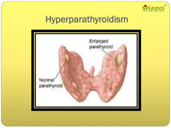
Hyperparathyroidism Nidal Younes MD Professor of Endocrine Surgery and Diabetic foot consultant- Jordan University Hospital
I.Hyperparatyhyroidism-Primary hyperparathyroidismTertiary HPT II. Malignancy-related-Solid tumor with metastases (breast)-Solid tumor with humoral mediation of hypercalcemia (lung, kidney)-Hematologic malignancies (multiple myeloma, lymphoma, leukemia) III. Endocrine diseases:Hyperthyroidism.Addisonian crisis.pheochromocytoma IV- Granulomatous diseases:Sarcoidosis.T.B. IV. Iatrogenic:Excessive intake of Vit D or calcium-Rx with lithium-Thiazide diuretics V. Associated with renal failure-Severe secondary hyperparathyroidism-Aluminum intoxication VI-Familial hypocalcuric hypercalcemia-Milk-alkali syndrome Hypercalcemia **Primary hyperparathyroidism and cancer account for 90% of cases of hypercalcemia
Incidence :0.1-0.3%. 1 case per 1000 men and 2-3 cases per 1000 women.25/100000 population Incidence increases above age 40 Most patients with sporadic PHPTarepostmenopausal women with an average age of 55 years Etiology: a solitary parathyroid adenoma(83%) Multiple adenomas (6%) Hyperplasia 10% Carcinoma 1% Primary Hyperparathyroidism
Primary HPT: Clinical Features • Symptomatic: • Classical pentad of symptoms(Kid.stones, • painful bones,abdominal groans,psychic moans,& fatigue overtones) • Osteitis fibrosa cystica • Nephrolithiasis • Pathologic fractures • Neuromuscular disease • Life-threatening hypercalcemia • DU.pancreatitis
Familial Syndromes • MEN I • MEN IIA • Familial Hypocalciuric Hypercalcemia • Hyperparathyroidism-jaw tumor syndrome • Fibro-osseous jaw tumors • Renal cysts • Solid renal tumors • Familial isolated hyperparathyroidism
MEN I • MEN I • 1 in 30,000 persons • Features: • Hyperparathyroidism (95%) • Most common and earliest endocrine manifestation • Gastrinoma (45%) • Pituitary tumor (25%) • Facial angiofibroma (85%) • Collagenoma (70%) • HPT in MEN I • Early onset • Multiple glands affected • Post-op hypoparathyroidism more common (more extensive surgery) • Successful subtotal parathyroidectomy followed by recurrent HPT in 10 years in 50% of cases
MEN IIA (Sipple’s Syndrome) • Features: • MTC(95%) • Pheochromocytoma(50%) • HPT(20%) • RET mutation (98%) • 1 in 30,000-50,000 people • Usually single adenoma but may have multi-gland hyperplasia
STIGMATA OF MEN I Lipomas Collagenomas Angiofibromas
This benign condition can be easily mistaken for mild hyperparathyroidism. It is an autosomal dominant inherited disorder characterized by hypocalciuria (usually < 50 mg/24 h), variable hypermagnesemia, and normal or minimally elevated levels of PTH. These patients do not normalize their hypercalcemia after subtotal parathyroid removal and should not be subjected to surgery. The condition has an excellent prognosis and is easily diagnosed with family history and urinary calcium clearance determination. Familial Hypocalciuric Hypercalcemia
Decreased GFR leads to reduced inorganic phosphate excretion and consequent phosphate retention Retained phosphate has a direct stimulatory effect on PTH synthesis and on cellular mass of the parathyroid glands Retained phosphate also causes excessive production and secretion of PTH through lowering of ionized Ca2+ and by suppression of calcitriol production Reduced calcitriol production results both from decreased synthesis due to reduced kidney mass and from hyperphosphatemia. Low calcitriol levels, in turn, lead to hyperparathyroidism via both direct and indirect mechanisms. Calcitriol is known to have a direct suppressive effect on PTH transcription and therefore reduced calcitriol in CRD causes elevated levels of PTH Reduced calcitriol leads to impaired Ca2+ absorption from the GI tract, thereby leading to hypocalcemia, which then increases PTH secretion and production. Secondary Hyperparathyroidism
Secondary HPT • Clinical presentation • Usually asymptomatic • Diagnosis • Elevated PTH in the setting of low or normal serum calcium is diagnostic • If phosphorous is elevated, cause is renal • If phosphorous is low, other causes of vit D deficiency should be sought • Prevention • Vit D replacement • Phosphorus binders [Sevelamer] • Treatment • Medical • Calcimimetic agents • Surgical • Considered in cases of refractory severe hypercalcemia, severe bone disease, severe pruritis, calciphylaxis, severe myopathy
Tertiary hyperparathyroidism develops in patients with long-standing secondary hyperparathyroidism, which stimulates the growth of an autonomous adenoma. A clue to the diagnosis of tertiary hyperparathyroidism is intractable hypercalcemia and/or an inability to control osteomalacia despite vitamin D therapy. Surgical Referral - calcium- phosphate product > 70 - severe bone disease and pain -intractable pruritus - extensive soft tissue calcification with tumoral calcinosis -calciphylaxis Tertiary Hyperparathyroidism
A35 year old patient had hypercalcemia, hypercalciuria, osteopenia and elevated PTH. In addition, there is a history of passing kidney stones
Tc-99Sestamibi parathyroid scan with single photon emission computerized tomography (SPECT) shows a bright signal representing a large parathyroid adenoma posterior to the right thyroid lobe. Pre-op serum Ca was 11.4 mg/dl and iPTH 208.4 pg/ml.