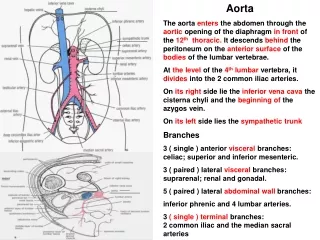
Slide 88, elastic artery (aorta), (10x obj.)
420 likes | 1.36k Vues
Slide 88, elastic artery (aorta), (10x obj.). TI. elastic fibers. TM. TA. Slide 36, elastic artery (aorta), trichrome (10x obj.). NOT vasa vasorum. TI. elastic fibers. TM. TA. elastic fibers. TM. vasa vasorum. TA. Slide 36, elastic artery (aorta), trichrome (40x obj.).

Slide 88, elastic artery (aorta), (10x obj.)
E N D
Presentation Transcript
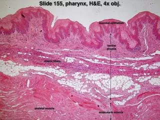
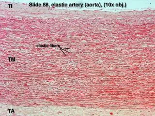
Slide 88, elastic artery (aorta), (10x obj.) TI elastic fibers TM TA
Slide 36, elastic artery (aorta), trichrome (10x obj.) NOT vasa vasorum TI elastic fibers TM TA
elastic fibers TM vasa vasorum TA Slide 36, elastic artery (aorta), trichrome (40x obj.)
Slide 30, muscular artery (40x obj.) • Vessel Tunics • A. Tunica Intima • endothelium • subendothelium • connective tissue • smooth muscle • (longitudinal fibers) • internal elastic lamina • B. Tunica Media • vascular smooth muscle • external elastic lamina • (circular fibers) • C. Tunica Adventitia • connective tissue • (longitudinal)
Slide 92, muscular artery w/ intimal thickening (40x obj.) • Vessel Tunics • A. Tunica Intima • endothelium • subendothelium • connective tissue • smooth muscle • (longitudinal fibers) • internal elastic lamina • B. Tunica Media • vascular smooth muscle • external elastic lamina • (circular fibers) • C. Tunica Adventitia • connective tissue • (longitudinal)
Slide 95, muscular artery, trichrome (40x obj.) internal elastic lamina external elastic lamina tunica media tunica adventitia elastic fibers
Slide 95, medium vein, H&E (10x obj.) • Tunica adventitia • thicker than tunica media • larger veins have longitudinal smooth muscle in adventitia • Tunica media • 3-15 layers of VSMCs • no external elastic lamina • Tunica intima • endothelium • subendothelium • IEL generally not evident
tunica adventitia tunica media Slide 95, medium vein, trichrome (4x obj.)
Slide 95, medium vein, trichrome (10x obj.) elastic fibers Note: IEL sometimes evident w/ trichrome stain tunica media tunica adventitia
Slide 95, arteriole and venule, H&E (40x obj.) • Arterioles • Tunica intima • very thin subendothelial layer • generally lacking distinct internal elastic lamina • Tunica media • 1-3 layers of VSMCs • no external elastic lamina • Tunica adventitia • thin and ill-defined • Venules • Tunica intima • endothelium • pericytes • Tunica media • 1-2 sparse layers of VSMCs • no external elastic lamina • Tunica adventitia • thicker than tunica media
Slide 95, arterioles and venules, trichrome (40x obj.) venule arteriole
Slide 95, arteriole and venule (?), trichrome (40x obj.) venule? arteriole? Remember, the change in vessel size is gradual, so you’ll find intermediate sizes. To make classification easy, focus on vessels that are clearly one type or another.
Slide 95, arteriole and venule (?), trichrome (40x obj.) arteriole? (sectioned obliquely) venule?
Slide 95, small vein & artery, H&E (40x obj.) vein artery
Slide 95, small vein & artery, trichrome (40x obj.) vein artery
valve Note number of white blood cells in lumen Slide 30, lymphatic vessel (10x obj.)
Slide 40, trachea (10x obj.) Look in this layer for arterioles and venules
Slide 40, trachea (40x obj.) venule arteriole
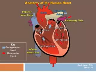
Slide 98, H&E (orientation) Look under endocardium to find Purkinje fibers chorda tendinae A-V valve leaflet papillary muscle atrial myocardium ventricular myocardium coronary vessels epicardium
Slide 98, H&E, (20x obj.) endocardium Purkinje fibers in “subendocardium” (note perinuclear space and large fiber size) myocardium cardiac muscle
Slide 98, H&E (orientation) Look under endocardium to find Purkinje fibers chorda tendinae A-V valve leaflet papillary muscle atrial myocardium ventricular myocardium coronary vessels epicardium
Slide 98, trichrome (20x obj.) endocardium Purkinje fibers (note perinuclear space and large fiber size) cardiac muscle
chorda tendinae papillary muscle A-V valve leaflet Note that distribution of elastic fibers faces blood flow. to atrium to ventricle Slide 98, trichrome (20x obj.)
Slide 99, H&E (orientation) aortic valve leaflet cardiac skeleton ventricular myocardium atrial myocardium A-V valve leaflet look here for A-V bundle of His
Slide 99, H&E (4x obj.) ventricular myocardium A-V bundle of His cardiac skeleton
Slide 99, H&E (20x obj.) A-V bundle of His cardiac skeleton
Slide 99, trichrome (orientation) aortic valve leaflet cardiac skeleton ventricular myocardium atrial myocardium A-V valve leaflet look here for A-V bundle of His
ventricular myocardium cardiac skeleton A-V bundle of His Slide 99, trichrome (4x obj.)
Slide 99, trichrome (20x obj.) A-V bundle of His cardiac skeleton
Slide 100, H&E (orientation) aortic valve leaflet ventricular myocardium atrial myocardium
Slide 100, Weigert (orientation) aortic valve leaflet ventricular myocardium atrial myocardium
Slide 100, Weigert (4x obj) to aorta aortic valve leaflet Note that distribution of elastic fibers faces blood flow. to ventricle
Slide 100, Weigert (4x obj) Note that distribution of elastic fibers faces blood flow. aortic valve leaflet aortic side of leaflet ventricular side of leaflet
Slide 102, H&E (orientation) coronary vessels epicardium ventricular myocardium atrial myocardium aortic valve leaflet