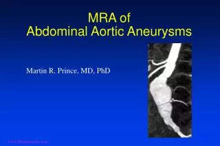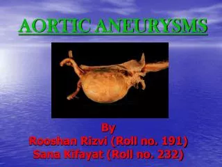
Aortic Aneurysms
Aortic Aneurysms. Dilshan Udayasiri. Some Anatomy. ascending aorta arch of the aorta descending aorta abdominal aorta. Layers of the aorta. Types of aneurysms. Shape. Location Thoracic (25%) Ascending (60%) Aortic Arch - includes brachiocephalic arteries (10% Descending (40%)

Aortic Aneurysms
E N D
Presentation Transcript
Aortic Aneurysms Dilshan Udayasiri
Some Anatomy ascending aorta arch of the aorta descending aorta abdominal aorta
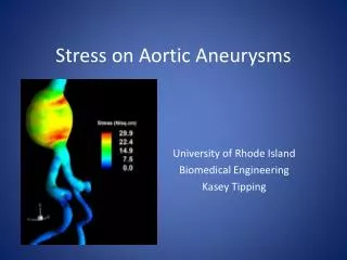
Types of aneurysms Shape • Location • Thoracic (25%) • Ascending (60%) • Aortic Arch - includes brachiocephalic arteries (10% • Descending (40%) • Thoracoabdominal (10%) • Abdominal (75%) Causes Degenerative Dissecting Saccular Fusiform Ruptured
Risk Factors • Hypertension • Hypercholesterolaemia • Smoking • Age (rare before 60) • Genetic (Marfans, Ehlers-Danlos syndrome) • Bicuspid Aortic Valve • Inflammatory/infectious - eg Giant Cell Arteritis
Symptoms • Incidental • Pain - tearing, radiating to back • Heart failure - due to AR • Thromboembolic (stroke, painful/parathesia of limbs) • Hoarseness of voice (compression of recurrent laryngeal nerve ) • Can mimic other acute disorders (AMI, renal colic, pancreatitis)
Signs • obs • lack of peripheral pulses • Pulsatile mass and tender abdomen • Murmur • Decreased BS and dullness to percussion • Signs of heart failure • Neurologic signs (Horner’s Syndrome - compression of cervical sympathetic ganglion)
Treatment • Watchful Waiting + medical • Percutaneous or open intervention
Watchful Waiting • Tight blood pressure control (MAP between 60 - 75) • beta blocker favourable unless contraindicated • persistent hypertension, check kidneys • cease smoking • treat hypercholesterolaemia • Screening • 6 months after initial scan then every 12 months unless symptomatic or increased rate of expansion or if size is 4.5cm - 5.5cm.
Indications for surgery • HD unstable • symptomatic • diameter ≥ 5.5cm • rate of growth ≥ 1.0cm/year
Endovascular repair Indications High perioperative risk pt’s Other Benefits shorter ICU stay Shorter Hospital Stay Quicker return to normal function Increased surviability in the short term Complications Endoleak (Type 1-4) Device Migration Infection Haematoma Stroke AMI Death
Surgery • Incision depends on location • Median sternotomy - arch • left thoracomtomy - descending • left thoracotomy extending across costal margin for retroperitoneal approach - thoracoabdominal • Abdominal incision - AAA Considerations Distal perfusion cerebral protection Renal Dysfunction Staged procedure