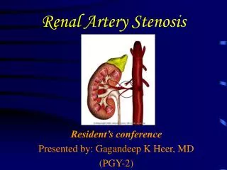
Renal Artery Stenosis
420 likes | 964 Vues
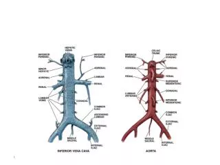
Renal Artery Stenosis. Case Studies. Coronal view of the proximal right RA (notice the high color velocity). Case #1= 52 yr. old male with severe HTN. IVC. Aorta. Case #1. Not a true peak velocity. Duplex scale is set too low ( 1.0 m/sec ). There is a musical bruit from a tight stenosis.

Renal Artery Stenosis
E N D
Presentation Transcript
Renal Artery Stenosis Case Studies
Coronal view of the proximal right RA (notice the high color velocity) Case #1= 52 yr. old male with severe HTN IVC Aorta
Case #1 Not a true peak velocity. Duplex scale is set too low ( 1.0 m/sec ). There is a musical bruit from a tight stenosis.
Case #1 True peak velocity. Duplex scale set properly ( 4.0 m/s ).
Case #1 Tardus-parvus seen in the segmental artery, indicating a proximal tight stenosis.
Case #1 Right renal artery pre-angioplasty, you can see the tight stenosis involving the proximal right renal artery ( arrow ).
Case #1 Right renal artery post angioplasty , the stenosis has resolved.
Case #1 Same right renal artery post-angioplasty.
Case #1 Normal color velocity in the right renal artery after angioplasty ( arrow ).
Case #1 Velocity returns to normal.
Case #1 Segmental waveform returns to normal .
Case # 2 : 40 year old male with HTN This patient had normal velocities in the proximal renal arteries bilaterally. But, the mid and distal renal arteries, both right and left had very high velocities, with tardus-parvus seen in the segmental arteries.
Case # 2 : 40 year old male with HTN Distal RRA
Case #2 Rounded waveforms in the segmental right renal artery Segmental RRA
Case #2 High velocities seen in the mid left renal artery Mid LRA
Case #2 High velocities seen in the distal left renal artery Distal LRA
Case #2 : RAR calculation Ultrasound worksheet: RAR in the Mid and Distal renal arteries are above 3.5 bilaterally, along with velocities above 180 cm/s, indicating a stenosis above 60%.
Case #3: Fibromuscular Dysplasia • Typical Angiographic appearance: • Beaded appearance of vessels • Mid and distal segments affected only. • This is where the ultrasound showed high velocities
Case #3 Post angioplasty. The arteries now have a smooth appearance. The blood pressure also returned to normal.
Case #4 72 year old female with HTN and increased BUN & Creatinine. Rt >2.8 cm than Lt.
Case #4 Proximal right RA with increased velocity.
Case #4 Abnormal Mid right renal artery
Case #4 Proximal left renal artery.
Case #4 Mid left renal artery. Notice the blunting of the mid artery’s waveform.
Case #4 Segmental left renal artery, has blunted waveform also.
Case #4 Angio confirms ultrasound findings of a bilateral high grade stenosis. The left renal artery is severely diseased. Left renal artery
Case #5: Elderly male with HTN Proximal right renal artery with elevated velocity.
Case #5 Segmental artery with Tardus-Parvus. Notice abnormal acceleration index.
Case #4 Post stent placement in the right artery. Left renal artery too diseased to stent.
Case #5 Example of a high grade stenosis with a normal segmental waveform. (absent Tardus-parvus). Remember not ALWAYS present in patients with a renal artery stenosis.
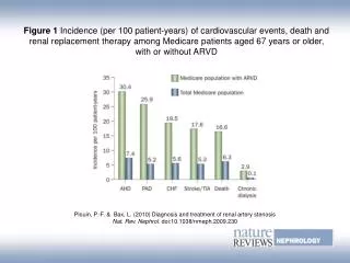
Summary Although Angiography is still considered the gold standard, Renal Artery Doppler is a very useful noninvasive screening test for renal artery stenosis. If US suggests RAS, PT usually have Angiogram for confirmation and possible treatment. If US normal, PT usually does not require further testing, sparing PT invasive risk and cost.