Download
1 / 12
160 likes | 978 Vues
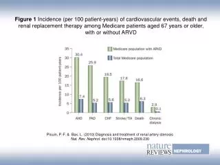
Pulmonic Stenosis & Aberrant Coronary Artery in an English Bulldog. Accession numbers 104145 and105676 Christina Copple, DVM Radiology Resident NCSU CVM-VTH. WINSTON. 2yr old M English Bulldog

E N D
Pulmonic Stenosis & Aberrant Coronary Artery in an English Bulldog Accession numbers 104145 and105676 Christina Copple, DVM Radiology Resident NCSU CVM-VTH
WINSTON • 2yr old M English Bulldog • August 2008: RDVM for lethargy and a distended abdomen with tachycardia, no episodes of collapse or coughing, no murmur was heard at this time. ECG revealed a sinus tachycardia • RDVM thoracic radiographs: cardiomegaly, possible pulmonary edema, and ascites • Started on Lasix - improved and became more active, abdominal distention decreased post Lasix http://www.the-bulldog.com/en/bull-dog-health/bulldog-news-longevity-of-british-breeds-of-dog.html
WINSTON • 1 month later NCSU Cardiology service • III/VI left basilar murmur, increased bronchovesicular sounds heard in all lung fields, pendulous and distended abdomen with palpable fluid wave • Diagnostics performed: thoracic radiographs, echocardiogram, ECG, and abdominocentesis
Pulmonic Stenosis (PS) • 3rd most common canine congenital cardiac defect • Sub-, supra- and primary valvular forms • Most common form of PS is primary dysplasia of the pulmonary valve http://www.the-bulldog.com/en/bull-dog-health/bulldog-news-longevity-of-british-breeds-of-dog.html
http://www.marvistavet.com/assets/images/pulmonary_stenosis.gifhttp://www.marvistavet.com/assets/images/pulmonary_stenosis.gif
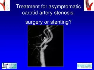
Diagnosis of PS • Suspect based on clinical signs, PE findings and breed • Thoracic radiographs • Echocardiogram • Angiography http://vmthpub.vetmed.wisc.edu/sa_services/med/cardiology/ultrasound.htm&usg
Treatment of PS • *If overt congestive heart failure has developed then chance of success is decreased • Mild to moderate, uncomplicated PS – no tx • Severe (systolic stenotic gradient of 100-125 mm Hg) or symptomatic PS candidates for balloon valvuloplasty or surgery (open patch-graft)
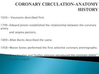
PS and Type R2A anomaly • Unique form of subvalvular PS • Left/Right coronary arteries originate from single right aortic sinus of valsalva • Compression of RVOT by encircling anomalous left coronary artery Normal Canine Angiogram http://www.vin.com/Members/CMS/Misc/default.aspx?id=8641
PS and Type R2A anomaly Accession # 105676 http://www.vin.com/Members/CMS/Misc/default.aspx?id=8641
Pathogenesis of Single Right Coronary Artery and Pulmonic Stenosis in English BulldogsBuchanan, James W. JVetMed. 2001 • Histology - failure of development of left coronary anlagen results in malformation of left aortic sinus and inversion of left main coronary artery • Leads to development of single right coronary artery and persistence of embryonic circulopulmonary conal artery (circle of Vieussens or Vieussen’s ring), which surrounds the pulmonary outflow tract Histologic cross-section at the level of the aortic sinuses of Valsalva in a stillborn dog with single right CA and subvalvular pulmonic stenosis. The right coronary sinus (R) and noncoronary sinus (N) are slightly larger than the left sinus and appear normal except for increased thickness of the wall of the right sinus and increased diameter of the right CA. In the left sinus, notice the inward protrusion of elastic media (arrows) at a point where the left CA should have extended outward from the aorta (A). Pulmonary valves (P). Right Atrium (RA). Left Atrium (LA). Van Gieson stain 20X. Bar = 1mm.
REFERENCES • Ettinger, Stephen J., and Edward C. Feldman. Textbook of Veterinary Internal Medicine. Volume II. Elsevier. St. Louis, MO. 2005, pp 998-1008. • Buchanan, James W. Pathogenesis of Single Right Coronary Artery and Pulmonic Stenosis in English Bulldogs. JVetInternMed. 2001; 15: 101-104. • Thrall, Donald E., DVM, PhD. Textbook of Veterinary Diagnostic Radiology. 5th ed. Saunders. St. Louis, MO. 2007. • Buchanan, James, DVM. Single Coronary Type R2A. James Buchanan Cardiology Library. Veterinary Information Network. http://www.vin.com/Members/CMS/Misc/default.aspx?id=8641.