Enteropathic Arthritis
Enteropathic Arthritis. Outline. Definition History Gut-synovium axis Enteropathic arthritis: Inflammatory Bowel Disease Mechanism Infectious enteritis (reactive arthritis) Whipple’s disease Intestinal bypass surgery Celiac disease. Definition.

Enteropathic Arthritis
E N D
Presentation Transcript
Outline • Definition • History • Gut-synovium axis • Enteropathic arthritis: • Inflammatory Bowel Disease • Mechanism • Infectious enteritis (reactive arthritis) • Whipple’s disease • Intestinal bypass surgery • Celiac disease
Definition • Arthropathies associated with disease of large and small intestines: • Inflammatory bowel disease • Crohn’s disease • Ulcerative colitis • Infectious enteritis (reactive arthritis) • Whipple’s disease • Intestinal bypass surgery • Celiac disease
History • 1922: Smith performed segmental bowel surgery to treat patients with RA • 1935: Hench describes arthritis flares during colitis exacerbation subsiding with remission • 1964: American Rheumatism Association considers enteropathic arthritis its own entity • 1976: Moll and Wright propose inclusion of the enteropathic groups into the seronegative sponyloarthropathies
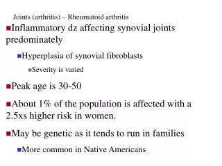
Patterns of Intestinal Inflammation in SpA Peng S. WRAMC. Feb 2009
Inflammation • Most with spondyloarthropathies do not have signs or symptoms of intestinal inflammation • Many have subclinical intestinal inflammation • 67% have macroscopic and microscopic gut inflammation on colonoscopy • Acute changes similar to infectious enterocolitis • Chronic changes suggestive of early CD • Chronic lesions more common with a family history of AS or CD Holden w, et al. Rheumatic Disease Clinics of North America 2003;29:513-530.
Inflammation De Vos, et al. Gastroenterology 1996;110(6):1696-703. • Acute inflammatory lesions • Normal mucosal structure with infiltration of epithelium with: • Neutrophils and eosinophils • Crypt abscess formation • Infiltration of lamina propria with PMN cells • Chronic inflammatory lesions • Disturbed mucosal architecture • Irregular, blunted fused villi • Distorted crypts and basal lymphoid aggregates
Inflammation • Reactive Arthritis • Acute lesions • Undifferentiated arthritis • Chronic > acute lesions • AS • Chronic >>acute lesions • Arthritis remission = normal gut histology • Joint flares = gut inflammation De Vos et al. Gastroenterol. 1996;110:1696. Mielants et al. J Rheumatol. 1995;22:2279.
Inflammation Rudwaleit M and Baeten D. Best Pract Res Clin Rheumatol. 2006;20:451-471.
Spondyloarthopathic Ileitis and Crohn’s Disease • Subclinical vs. preclinical Crohn’s disease • Study of 123 patients with SpA who underwent ileocolonoscopy • Baseline and repeat at 2-9 years • Normal 32% • Acute lesions 23 % • Chronic lesions 45% 6% developed CD Rudwaleit M and Baeten D. Best Pract Res Clin Rheumatol. 2006;20:451-471
Risk Factors for Arthritis and GI Disease • Chronic inflammation at greatest risk • Peristently high C-reactive protein • Radiographic sacroiliitis in the absence of HLA-B27
Inflammatory Bowel Disease • Crohn’s: • Entire GI tract from mouth to anus: • Ileitis 30% • Ileocolitis 40% • Colitis 30% • Bimodal age distribution • 20-100 per 100,000 • Skip Lesions and transmural inflammation
Ulcerative Colitis • Limited to the colon, most have rectal involvement • Proctosigmoiditis 50% • Left sided colitis 30% • Extensive colitis 20% • Age 20-25 yrs • 70-150 per 100,000 • Contiguous lesions • Micro-ulcerations • Crypt abscesses
IBD and Peripheral Arthritis • 9-53% of patients with IBD • Arthritis is the most common extra-intestinal manifestation of IBD • More likely with large bowel disease • Can occur before bowel symptoms and at any time in the disease course • Most frequent with extensive UC and CD: • Abscesses • Perianal disease • Erythema Nodosum • Pyoderma Gangrenosum
IBD and Peripheral Arthritis • Male=Female • Occurs with children and adults • Relationship between flares and severity of bowel disease (UC) • In UC, surgical resection of diseased segment usually stops the arthritis • In CD, surgical resection does not help arthritis
Classification of Arthritis • Peripheral arthritis: • Type I • Pauciarticular (5 or fewer joints) • Type II • Polyarticular • Axial Arthritis (Type III): • Sacroiliitis/Spondylitis
Type 1 (Pauciarticular) Peripheral Arthritis • Equal incidence between males and females • Peak age of onset 25-44 • Incidence is 3.6% in UC and 6% in Crohn’s • Often parallels the intestinal activity • Associated with HLA-DRB1*0103, HLA-B27 and HLA-B*35 Orchard TR,et al. Gut 1998;42:387-391.
Type 1 (Pauciarticular) Peripheral Arthritis Pauciarticular arthritis: • Less than 5 joints • Most common joint: knee • Acute (< 10 weeks) • Self-limiting 90% less than 6 months • Associated with IBD flares • Strongly associated with extra-intestinal manifestations of IBD such as uveitis and EN • Holden w, et al. Rheumatic Disease Clinics of North America 2003;29:513-530.
Type II (Polyarticular) Peripheral Arthritis • Incidence: • 2.5% in UC and 4% in Crohn’s • Polyarticular arthritis: • 5 or more joints • Most common: MCP’s • No HLA-B27 association • Associated with HLA-B*44 • Exacerbations/remissions
Type II (Polyarticular) Peripheral Arthritis • Chronic course independent of activity of the IBD • Arthritis activity does not parallel bowel activity • Duration: persists for months to years • Associated with uveitis but not with other extraintestinal manifestations
Peripheral Arthritis • Peripheral arthritis associated with IBD is seronegative • Typically non-deforming and nonerosive • Erosive disease affecting the hips, elbows, MCP joints, MTP joint and erosive polyarthritis has been described • In MCP and MTP’s, arthropathy differs from RA as it is predominately asymmetric
Axial (Type 3) Arthritis • Ulcerative colitis 2-6%, Crohn’s 5-22% • Presents as either: • Spondylitis: • 1-26% of IBD pts • May occur with type I arthritis • Sacroiliitis: • Asymptomatic • 4-18% of IBD pts • Ankylosing spondylitis-like • 3-10% of IBD pts
Axial (Type 3) Arthritis • Male: female ratio 2:1 • Occurs at any age • Axial involvement and IBD course are usually independent • Usually precede onset of IBD by many years • Genetic associations • CARD15 and ? HLA-B27 • Inflammatory back pain • Lumbar straightening, dorsal kyphosis, limited chest expansion
Genetics and IBD • Concordance in extra-intestinal manifestations of IBD • 70% in parent-child pairs • 84% in sibling pairs • HLA-B27 • CARD 15 • Others: NOD2, HLA-B35, HLA-DRB1*0103, HLA-B44
HLA-B27 • Pathologic role unknown • Impaired ability to process/present bacterial antigen to immune cells • Constant source of immune stimulation • Strongest association with idiopathic AS • Less crucial in patients with IBD • 50-70% + for HLA-B27
CARD 15 • Gene coding for a protein involved in innate immunity • Increased risk of Crohn’s disease • 78% of Crohn’s patients with sacroiliitis • 48% of Crohn’s patients without sacroiliitis • Association is independent of HLA-B27
Rudwaleit M, et al. Best Pract Res Clin Rheumatol. 2006;20:451-471.
Mechanism • Breach in GI wall integrity • Increased permeability to macromolecules • Increased exposure to microbial and dietary antigens • Loss of tolerance to own bacterial flora • Host susceptibility to the increased antigenic load • Recirculation of antigen-specific memory T-cells from gut to joints
Laboratory • Lab findings as determined by activity of IBD • IDA, leukocytosis, thrombocytosis common • RF negative • Acute phase reactants increased • HLA-B27and other HLA typing • Synovial fluid: WBC 1500 – 50,000 • PMN predominate • Synovial membrane biopsy: • Mild chronic inflammation indistinguishable from RA • Proliferation of synovial lining cells, increased vascularity, infiltration of mononuclear cells
Radiology • SI joints • Indistinguishable from AS • Bilateral sacroiliac erosions • “Pseudowidening” of the SI joint • Fusion with complete obliteration of SI joint • MRI is most sensitive/specific for sacroiliitis
Radiology • Spine: • Shiny corners or Romanus lesions • Syndesmophytes • Symmetric, delicate appearing, marginal • Peripheral joints: • Soft tissue swelling, juxta-articular osteoporosis, mild periositis, effusion • Usually without erosions/destructions • Enthesitis: • Faint periosteal reaction at bony prominence
Ocular Manifestations • Anterior Uveitis: 0.5-3% of pts with IBD • Unilateral vs. bilateral • Associated with HLA-B27 and axial involvement • Painful red eye, blurred vision and photophobia • Optho consult and therapy with topical/systemic CS • Episcleritis: 2-5% • Hyperemia of sclera and conjunctiva • No vision loss, painless • TX: • Treat underlying IBD • NSAIDs and topical steroids www.uptodate.com
Skin Manifestations • Erythema Nodosum: 10-15% • Raised, warm tender nodules • Coincides with exacerbations of IBD and thus with peripheral arthritis • Therapy: NSAIDs, colchicine, TNF-inhibitors • Pyoderma Gangrenosum: • More common in UC 5% • Associated with severe IBD • Therapy: • Systemic CS • Infliximab, cyclosporine, cellcept www.nature.com
Osteoporosis • Prevalence of 15% • RR for fracture in IBD is 40-60% • Inflammatory bone-resportive cytokines • IL-1,IL-6 and activated T cells • Higher levels of RANKL expressed in CD • Therapy: • Bisphosphonates • Calcium/vitamin D • Minimize steroid use • Weight bearing exercises Kethu S. J Clin Gastroenterol 2006; 40(6):467-475.
Therapy • NSAIDS: • Adverse effects on bowel symptoms • Concern about NSAID’s and development of IBD • Limited evaluation with COX-2 inhibitors • Sulfasalazine • Azathioprine • 6-mercaptopurine • Aminosalicylates (mesalamine)
Therapy • Biologics: • Infliximab • Beneficial for Crohn’s disease • Fistula formation • Perianal disease • Less efficacy in UC • Etanercept • No benefit for GI disease • Adalimumab • Approved for AS and IBD • Similar benefits to infliximab
Therapy • Type 1 peripheral arthritis: • Treat the IBD • Sulfasalazine, MTX, AZA, TNF inhibitors and CS • Type II and III (axial) arthritis • No improvement with medical or surgical treatment of IBD • NSAIDs effective but may exacerbate gut symptoms • Sulfasalazine, MTX and Azathioprine • TNF inhibitor therapy: use after no response with NSAIDs at 3 months
Rudwaleit M and Baeten D. Best Pract Res Clin Rheumatol. 2006;20:451-471
Enteropathic Enteropathy • Reactive Arthritis (Infectious enteritis) • Whipple’s • Intestinal Bypass surgery • Celiac Disease
Whipple’s Disease • Rare multisystem systemic disease • Infection caused by Tropheryma Whippelii • Predominance in the small bowl • Male to female ratio of 9:1 • Mean age 55 (range 20-82) • Symptoms: • Diarrhea, weight loss, fever and arthritis • LAD, hyperpigmentation, serositis, CNS
Whipple’s disease • Lab abnormalities: • Anemia, hypoalbuminemia, low serum carotene and iron • Increased stool fat • Biopsy: • Villi become distended with foamy and PAS + macrophages • Rod-shaped free bacilli in the lamina propria • PCR of tissue or blood
Whipple’s disease www.uptodate.com
Whipple’s disease • Peripheral arthritis • Polyarticular and symmetric • 67% as their only symptoms • Migratory and episodic • Precedes GI symptoms by 5 years • Arthritis flares not related to GI symptoms • Erosions rare • Axial arthritis • Incidence controversial 8-20% • Relation to HLA-B27 unclear
Treatment • Fatal if not treated • Initial antibiotic therapy: • Ceftriaxone 2 grams IV q 12+ streptomycin 1 g IV q 24 hrs for 10-14 days • Then one year of • Trimethoprim-sulfamethoxazole : 1 DS po bid • Doxycycline: 100 mg po bid • Can see Jarisch-Herxheimer reaction • Fevers, chills, headache, hypotension, severe abdominal pain or pleuritic chest pain
Intestinal bypass arthritis • Jejunocolic and jejunoileal bypass surgeries • Inflammatory joints in 6-52% • Usually within 3 years of surgery • Females > males • No HLA association • Arthritis: • Peripheral symmetric, polyarticular, • Knees, wrists, MCP’s and MTP’s • Vesiculopustular skin rash • Occurs in over ½ of patients with arthritis
Intestinal bypass arthritis • Pathogenesis: • Bacterial overgrowth in blind loop • Mucosal changes allow increased absorption of bacterial antigens • Formulation and circulation of immune complexes • Immune complexes demonstrated in synovium, synovial fluid and skin lesions