Download
1 / 36
360 likes | 525 Vues
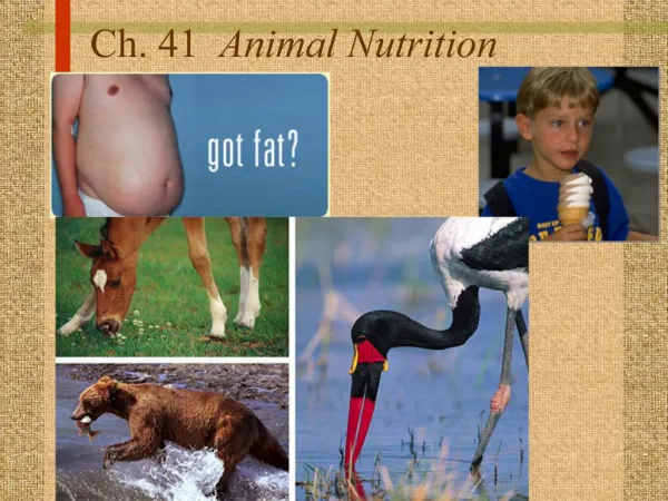
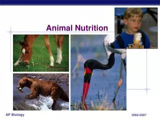
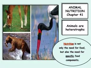
Ch.41 Animal Nutrition. Everyone has to eat. What tells you, you are hungry?. A. Nutritional Requirements of Animals 1. Homeostatic mechanisms manage an animal’s fuel.

E N D
A. Nutritional Requirements of Animals • 1. Homeostatic mechanisms manage an animal’s fuel. • Nearly all ATP generation is based on the oxidation of organic fuel molecules—carbohydrates, proteins, and fats—in cellular respiration. • Fats are rich in energy, liberating about twice the energy from an equal amount of carbohydrate or protein during oxidation. • In humans, the liver and muscle cells store energy as glycogen, a polymer made up of many glucose units. • If glycogen stores are full and caloric intake still exceeds caloric expenditure, the excess is usually stored as fat. • When fewer calories are taken in than are expended—perhaps because of sustained heavy exercise or lack of food—fuel is taken out of storage depots and oxidized. • The human body expends liver glycogen first and then draws on muscle glycogen and fat. • Most healthy people—even if they are not obese—have enough stored fat to sustain them through several weeks of starvation. • If the diet of a person or other animal is chronically deficient in calories, undernourishment results. • The stores of glycogen and fat are used up, the body begins breaking down its own proteins for fuel, muscles begin to decrease in size, and the brain can become protein-deficient.
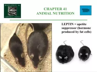
2. Obesity is a global health problem. • Overnourishment, or obesity, the result of excessive food intake, is a common problem in the United States and other affluent nations. • The human body tends to store any excess fat molecules obtained from food instead of using them for fuel. • In contrast, when we eat an excess of carbohydrates, the body tends to increase its rate of carbohydrate oxidation. • Thus, the amount of fat in the diet can have a more direct effect on weight gain. • In the United States, the percentage of obese people has doubled to 30% over the past 20 years, and another 35% are overweight. • Obesity contributes to health problems, including diabetes, cancer of the colon and breast, and cardiovascular disease. • In mammals, a hormone called leptin, produced by adipose cells, is a key player in a complex feedback mechanism regulating fat storage and use.
As adipose tissue increases, high leptin levels cue the brain to depress appetite and to increase energy-consuming muscular activity and body-heat production. • Conversely, loss of body fat decreases leptin levels in the blood, signaling the brain to increase appetite and weight gain. • For some reason, the brain’s satiety center does not respond to the high leptin levels in many obese people. • One hypothesis is that in humans, in contrast to other mammals, the leptin system functions to stimulate appetite and prevent weight loss rather than to prevent weight gain. • Most humans crave fatty foods. Although fat hoarding is a health liability today, it may have been advantageous in our evolutionary past. • Natural selection may have favored those individuals with a physiology that induced them to gorge on fatty foods on the rare occasions that they were available.
3. An animal’s diet must supply essential nutrients and carbon skeletons for biosynthesis. • Besides fuel and carbon skeletons, an animal’s diet must also supply essential nutrients. • An animal whose diet is missing one or more essential nutrients is said to be malnourished. • Malnutrition is much more common than undernourishment in human populations, and it is even possible for an overnourished individual to be malnourished. • There are four classes of essential nutrients: essential amino acids, essential fatty acids, vitamins, and minerals. • Animals require 20 amino acids to make proteins. • Eight amino acids are essential in the adult human with a ninth, histidine, being essential for infants. • A diet that provides insufficient amounts of one or more essential amino acids causes a form of malnutrition known as protein deficiency. • In one variation of protein malnutrition, called kwashiorkor, the diet provides enough calories but is severely deficient in protein.
The protein in animal products, such as meat, eggs, and cheese, are “complete,” which means that they provide all the essential amino acids in their proper proportions. • Most plant proteins are “incomplete,” being deficient in one or more essential amino acid. • Because the body cannot easily store amino acids, a diet with all essential amino acids must be eaten each day, or protein synthesis is retarded. • While animals can synthesize most of the fatty acids they need, they cannot synthesize essential fatty acids. • Vitamins are organic molecules required in the diet in quantities that are quite small compared with the relatively large quantities of essential amino acids and fatty acids animals need. • So far, 13 vitamins essential to humans have been identified. • These can be grouped into water-soluble vitamins and fat-soluble vitamins. • The water-soluble vitamins include the B complex, which consists of several compounds that function as coenzymes in key metabolic processes. • Vitamin C, also water soluble, is required for the production of connective tissue. • Excessive amounts of water-soluble vitamins are excreted in urine, and moderate overdoses are probably harmless. • The fat-soluble vitamins are A, D, E, and K. • Vitamin A is incorporated in the visual pigments of the eye. • Vitamin D aids in calcium absorption and bone formation. • Vitamin E seems to protect membrane phospholipids from oxidation. • Vitamin K is required for blood clotting. • Excess amounts of fat-soluble vitamins are not excreted but are deposited in body fat. • Overconsumption may lead to toxic accumulations of these compounds.
Minerals are simple inorganic nutrients, usually required in small amounts—from less than 1 mg to about 2,500 mg per day. • Humans and other vertebrates require relatively large quantities of calcium and phosphorus for the construction and maintenance of bone. • Calcium is also necessary for the normal functioning of nerves and muscles. • Phosphorus is a component of the cytochromes that function in cellular respiration. • Iron is a component of the cytochromes that function in cellular respiration and of hemoglobin, the oxygen-binding protein of red blood cells. • Magnesium, iron, zinc, copper, manganese, selenium, and molybdenum are cofactors built into the structure of certain enzymes. • Iodine is required for thyroid hormones, which regulate metabolic rate. • Sodium, potassium, and chloride are important in nerve function and have a major influence on the osmotic balance between cells and the interstitial fluids.
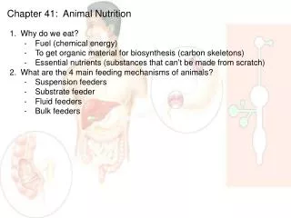
B. Overview of Food Processing • 1. The four main stages of food processing are ingestion, digestion, absorption, and elimination. • Ingestion, the act of eating, is only the first stage of food processing. • Digestion, the second stage of food processing, is the process of breaking food down into molecules small enough for the body to absorb. • Digestion cleaves macromolecules into their component monomers, which the animal then uses to make its own molecules or as fuel for ATP production. • Polysaccharides and disaccharides are split into simple sugars. • Fats are digested to glycerol and fatty acids. • Proteins are broken down into amino acids. • Nucleic acids are cleaved into nucleotides. • Digestion breaks bonds with the addition of water via enzymatic hydrolysis. • After the food is digested, the animal’s cells take up small molecules such as amino acids and simple sugars from the digestive compartment, a process called absorption. • During elimination, undigested material passes out of the digestive compartment. • 2. Digestion occurs in specialized compartments.
C. The Mammalian Digestive System • Peristalsis, rhythmic waves of contraction by smooth muscles in the walls of the canal, pushes food along. • Sphincters, muscular ringlike valves, regulate the passage of material between specialized chambers of the canal. • The accessory glands include the salivary glands, the pancreas, the liver, and the gallbladder.
1. The oral cavity, pharynx, and esophagus initiate food processing. • Both physical and chemical digestion of food begins in the mouth. • The presence of food in the oral cavity triggers a nervous reflex that causes the salivary glands to deliver saliva through ducts to the oral cavity. • Chemical digestion of carbohydrates, a main source of chemical energy, begins in the oral cavity. • Saliva contains salivary amylase, an enzyme that hydrolyzes starch and glycogen into smaller polysaccharides and the disaccharide maltose. • The tongue tastes food, manipulates it during chewing, and helps shape the food into a ball called a bolus. • The pharynx, also called the throat, is a junction that opens to both the esophagus and the trachea (windpipe). • When we swallow, the top of the windpipe moves up so that its opening, the glottis, is blocked by a cartilaginous flap, the epiglottis. • The esophageal sphincter relaxes and the bolus enters the esophagus. • In the meantime, the larynx moves downward and the trachea is opened, and peristalsis moves the bolus down the esophagus to the stomach. • The esophagus conducts food from the pharynx down to the stomach by peristalsis. • The muscles at the very top of the esophagus are striated and, therefore, under voluntary control. • Involuntary waves of contraction by smooth muscles in the rest of the esophagus then take over.
2. The stomach stores food and performs preliminary digestion. • The stomach is located in the upper abdominal cavity, just below the diaphragm. • With accordionlike folds and a very elastic wall, the stomach can stretch to accommodate about 2 L of food and fluid, storing an entire meal. • The stomach also secretes a digestive fluid called gastric juice and mixes this secretion with the food by the churning action of the smooth muscles in the stomach wall. • Also present in gastric juice is pepsin, an enzyme that begins the hydrolysis of proteins. • Pepsin, which works well in strongly acidic environments, breaks peptide bonds adjacent to specific amino acids, producing smaller polypeptides. • Pepsin is secreted in an inactive form called pepsinogen by specialized chief cells in gastric pits. • In a positive-feedback system, activated pepsin can activate more pepsinogen molecules. • The stomach’s second line of defense against self-digestion is a coating of mucus, secreted by epithelial cells, that protects the stomach lining. • About every 20 seconds, the stomach contents are mixed by the churning action of smooth muscles. • As a result of mixing and enzyme action, what begins in the stomach as a recently swallowed meal becomes a nutrient-rich broth known as acidchyme. • At the opening from the stomach to the small intestine is the pyloric sphincter, which helps regulate the passage of chyme into the intestine.
3. The small intestine is the major organ of digestion and absorption. • With a length of more than 6 m in humans, the small intestine is the longest section of the alimentary canal. • Most of the enzymatic hydrolysis of food macromolecules and most of the absorption of nutrients into the blood occurs in the small intestine. • In the first 25 cm or so of the small intestine, the duodenum, acid chyme from the stomach mixes with digestive juices from the pancreas, liver, gall bladder, and gland cells of the intestinal wall. • Pancreatic enzymes include protein-digesting enzymes (proteases) that are secreted into the duodenum in inactive form. • The liver performs a wide variety of important functions in the body, including the production of bile. • Bile is stored in the gallbladder until needed. • It contains bile salts that act as detergents that aid in the digestion and absorption of fats. • Bile also contains pigments that are by-products of red blood cell destruction in the liver. • These bile pigments are eliminated from the body with the feces. • Most digestion is completed while the chyme is still in the duodenum. • The remaining regions of the small intestine, the jejunum andileum, function mainly in the absorption of nutrients and water.
The small intestine has a huge surface area—300 m2, roughly the size of a tennis court. • Large circular folds in the lining bear fingerlike projections called villi, and each epithelial cell of a villus has many microscopic appendages called microvilli that are exposed to the intestinal lumen. • Fructose, a simple sugar, moves by diffusion alone down its concentration gradient from the lumen of the intestine into the epithelial cells and then into capillaries. • Amino acids and sugars pass through the epithelium, enter capillaries, and are carried away from the intestine by the bloodstream. • Glycerol and fatty acids absorbed by epithelial cells are recombined into fats. • The fats are mixed with cholesterol and coated with special proteins to form small globules called chylomicrons. • The capillaries and veins that drain nutrients away from the villi converge into the hepatic portal vein, which leads directly to the liver. • The liver modifies and regulates this varied mix before releasing materials back into the bloodstream.
4. Reclaiming water is a major function of the large intestine. • The large intestine, or colon, is connected to the small intestine at a T-shaped junction where a sphincter controls the movement of materials. • One arm of the T is a pouch called the cecum. • The relatively small cecum of humans has a fingerlike extension, the appendix, which makes a minor contribution to body defense. • A major function of the colon is to recover water that has entered the alimentary canal as the solvent to various digestive juices. • About 7 L of fluid are secreted into the lumen of the digestive tract of a person each day. • More than 90% of the water is reabsorbed, most in the small intestine, the rest in the colon. • Digestive wastes, the feces, become more solid as they are moved along the colon by peristalsis. • If the lining of the colon is irritated by a bacterial infection, less water than usual is resorbed, resulting in diarrhea. • If insufficient water is absorbed because peristalsis moves the feces too slowly, the result is constipation.
Living in the large intestine is a rich flora of mostly harmless bacteria. • One of the most common inhabitants of the human colon isEscherichia coli, a favorite research organism. • As a by-product of their metabolism, many colon bacteria generate gases, including methane and hydrogen sulfide. • Some bacteria produce vitamins, including biotin, folic acid, vitamin K, and several B vitamins, which supplement our diet. • Feces contain masses of bacteria and undigested materials including cellulose. • Although cellulose fibers have no caloric value to humans, their presence in the diet helps move food along the digestive tract. • The terminal portion of the colon is called the rectum, where feces are stored until they can be eliminated. • Between the rectum and the anus are two sphincters, one involuntary and one voluntary.
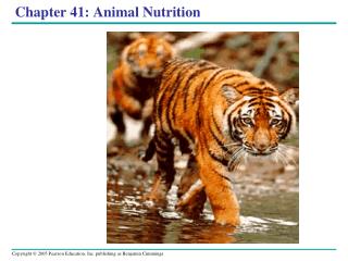
D. Evolutionary Adaptations of Vertebrate Digestive Systems • 1. Structural adaptations of digestive systems are often associated with diet. • Dentition, an animal’s assortment of teeth, is one example of structural variation reflecting diet. • For example, poisonous snakes, such as rattlesnakes, have fangs, modified teeth that inject venom into prey. • Large, expandable stomachs are common in carnivores, which may go for a long time between meals and, therefore, must eat as much as they can when they do catch prey. • For example, a 200-kg African lion can consume 40 kg of meat in one meal. • The length of the vertebrate digestive system is also correlated with diet. • In general, herbivores and omnivores have longer alimentary canals relative to their body sizes than do carnivores, providing more time for digestion and more surface areas for absorption of nutrients. • Vegetation is more difficult to digest than meat because it contains cells walls. • 2. Symbiotic microorganisms help nourish many vertebrates. • Much of the chemical energy in the diet of herbivorous animals is contained in the cellulose of plant cell walls. • However, animals do not produce enzymes that hydrolyze cellulose. • Many vertebrates (and termites) solve this problem by housing large populations of symbiotic bacteria and protists in special fermentation chambers in their alimentary canals. • These microorganisms do have enzymes that can digest cellulose to simple sugars that the animal can absorb. • Many herbivorous mammals, including horses, house symbiotic microorganisms in a large cecum, the pouch where the small and large intestines connect. • The symbiotic bacteria of rabbits and some rodents live in the large intestine and cecum. • The koala also has an enlarged cecum, where symbiotic bacteria ferment finely shredded eucalyptus leaves.
The most elaborate adaptations for a herbivorous diet have evolved in the ruminants, which include deer, cattle, and sheep. • When the cow first chews and swallows a mouthful of grass, boluses enter the rumen and the reticulum. • Symbiotic bacteria and protists digest this cellulose-rich meal, secreting fatty acids. • Periodically, the cow regurgitates and rechews the cud, which further breaks down the cellulose fibers. • The cow then reswallows the cud to the omasum, where water is removed. • The cud, with many microorganisms, passes to the abomasum for digestion by the cow’s enzymes.