LYMPHATIC SYSTEM
LYMPHATIC SYSTEM. Marieb - Chapter 20. Lymphatic System. capillaries ducts nodes lymphoid organs diffuse cell pockets. Lymphatic capillaries. extremely permeable present throughout body intermingle with blood capillaries not in CNS or mineralized tissues. Lymphatic Ducts. Trunks


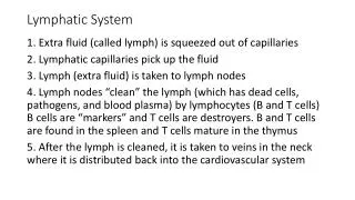
LYMPHATIC SYSTEM
E N D
Presentation Transcript
LYMPHATIC SYSTEM Marieb - Chapter 20
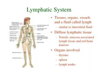
Lymphatic System • capillaries • ducts • nodes • lymphoid organs • diffuse cell pockets
Lymphatic capillaries • extremely permeable • present throughout body intermingle with blood capillaries • not in CNS or mineralized tissues
Lymphatic Ducts • Trunks • Lumbar • Bronchomediastinal • Subclavian • Jugular • Intestinal
Trunks anastomose & enter veinous return at junction of subclavian & jugular veins
Lymph Vessel Structure • much like veins • more valves • more frequent anastomoses
A tropical disease caused by lymphatic obstruction. Victim is bitten by a mosquito infected with a roundworm known as a filarial worm. The resulting edema leads to fibrosis and elephant-like thickening of the skin. ELEPHANTIASIS
Lymph Nodes • Encapsulated by dense fibrous connective tissue • Trabeculae - capsule extends inward form compartments
Lymph Nodes • Clusters at junctions of vessels into trunks
Lymph node structure Figure 20.4a • Cortex • dense follicles with germinal centers • dividing B cells • Deep cortex (T cells) • Medulla • inward extensions of cortical tissue (B, T and plasma cells) • Sinuses • lymph drainage • filtered by macrophages • Circulation in the node: • afferent lymphatic vessel subcapsular sinus drain through tissue hilus efferent vessels
Thymus • primary function is in early life • Secretes thymosin and thymopoietin which causes T lymphocytes to become immunocompetent • Lacks B cells (no follicles) • Atrophies with age: prominent in newborns, stops growth by adolescence, degenerates by old age
Lymphoid Follicle Aggregates • MALT (mucosa associated lymphatic tissue) includes: • Peyer’s Patches in intestines • Appendix • Tonsils • Small bronchiolar follicles • MALT are positioned well to: • Destroy bacteria that breach the mucosal membrane from outside • Develop “memory” lymphocytes for long term immunity
Tonsils • not encapsulated • blind pouches – crypts • bacteria can enter • induced immune response