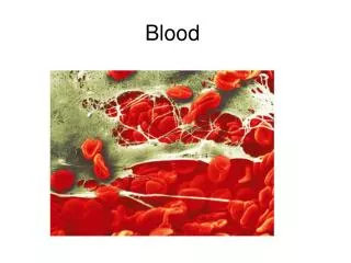
Blood
Blood. Chapter 17. Introduction. Circulatory System is subdivided into the: Cardiovascular system – blood, heart, and blood vessels Lympathetic system – lymph and lymph nodes and lymph vessels First we will look at blood – the river of life Hematology – the study of blood

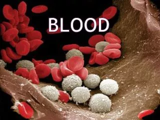
Blood
E N D
Presentation Transcript
Blood Chapter 17
Introduction Circulatory System is subdivided into the: • Cardiovascular system – blood, heart, and blood vessels • Lympathetic system – lymph and lymph nodes and lymph vessels First we will look at blood – the river of life • Hematology – the study of blood - clinicians examine it more often than any other tissue when investigating causes of disease in their patients
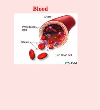
Blood Circulation • Initiated by the pumping action of the heart - blood leaves the heart in arteries tiny capillaries enter body tissues - from capillaries oxygen-deficient blood flows into veins which return it to the heart lungs heart Functions of blood: • Carries respiratory gases, nutrients, hormones • Conveys cells of the defense system to infectious sites • Helps regulate body temperature – blood is diverted to or away from the skin
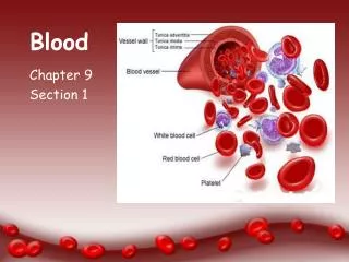
Overview: Composition of Blood • Blood accounts for ~8% of body mass – volume in male is 5 – 6 liters; females: 4 - 5 liters • A specialized Connective tissue - Blood cells (formed elements) are suspended in plasma (fluid portion and fibrinogen) • Hematocrit (‘blood fraction’) - % of the blood volume that consists of erythrocytes - males: 47 ± 5% - females: 42% ± 5%
Overview: Composition of Blood • Heavier formed elements are packed down (erythrocytes ‘red cells’) and the less dense plasma remains on top • Buffy coat junction between RBCs and plasma contains leukocytes (‘white cells’); platelets (thrombocytes) Figure 17.1
Blood Plasma • Straw-colored, sticky fluid portion of blood about 90% water Contains: • Ions – Na+ and Cl- • Nutrients – sugars, amino acids, lipids, • Wastes – urea and ammonia, and CO2 • O2, hormones and vitmins • 3 main types of proteins: - albumin helps to prevent water from diffusing out - globulins include both antibodies and blood proteins that transport lipids, iron, and copper - fibrinogens are invloved in blood clotting
Formed Elements Blood cells: • Erythrocytes lack nuclei and organelles • Platelets are cell fragments • Most cannot divide - survive for only a short time Staining of blood cells – cellular structures stain differentially according to their chemical makeup • Eosin an acidic dye – stains pink • Methylene blue a basic dye – stains blue and purple
Erythrocytes (RBCs) • Small, oxygen-transporting cells – 7.5 um in diameter (capillary diameter 8 – 10 um) - have no organelles or nuclei - cytoplasm packed with hemoglobin - generate energy by anaerobic mechanisms • Most numerous of the formed elements – females: 4.3 – 5.2 million cells/cubic ml of blood males: 5.1 – 5.8 million
An Erythrocyte • 30% more surface area for rapid diffusion of O2 • Sacs of hemoglobin (O2 and CO2) • Lives 100 -120 days • Originate in the bone marrow (expel their organelles) Figure 17.3
Leukocytes (WBCs) • Less numerous 4,800 – 11,000 per cubic ml - originate in the bone marrow and are released continuously into the blood • Protect the body from infectious microorganism • Function outside the blood stream in loose CT where infections occur • Diapedesis (‘leaping through’) – circulating leukocytes leave the capillaries by squeezing between endothelial cells of the capillary walls - travel by amoeboid motion to infection sites
WBCs • Bone marrow stores leukocytes -releases in large quantities during serious infections - count exceeding 11,000 per cubic ml = leukocytosis • 5 types divided into 2 groups (presence or absence of membrane-bound cytoplasmic granules): 1) Granulocytes – neutrophils, eosinophils, basophils 2) Agranulocytes – lymphocytes and monocytes Relative amount of leukocytes from most to least: Never Let Monkeys Eat Bananas
Relative Percentages of the Different Types of Leukocytes Figure 17.5
Granulocytes • Neutrophils – most numerous WBC (60%) • Phagocytize and destroy bacteria - also release bacteria-destroying substances into ECM of the infected tissue - pus is composed of dead neutrophils and other WBCs plus tissue debris and dead bacteria • Nucleus – 2 to 6 lobes • Granules pick up acidic and basic stains - membrane-walled sacs of digestive enzymes
Neutrophil • Nucleus – has two to six lobes • Granules pick up acidic and basic stains Figure 17.4a
Eosinophils: 1 – 4% of all WBCs that function during allergic reactions and parasitic infections (gather in digestive tube wall) • Granules contain digestive enzymes - phagocytose allergens then degrade histamine - attach to parasites release enzymes that digest and destroy the invaders Figure 17.4b
Figure 17.4c • Basophils (base loving) – rarest, about 0.5% of all leukocytes • Nucleus – usually two lobes • Granules secrete histamines and molecules • Function in inflammation mediation
Agranulocytes • Lymphocytes (T and B cells) – most important cells of the immune system (20 – 45% of WBCs) Nucleus – occupies most of the cell volume (stains dark purple) • Function in lymphoid CTs – play a crucial role in immunity - effective in fighting infectious organisms - act against antigens (‘induce against’) a specific foreign molecule that induces a response - T cells attack foreign cells directly - B cells differentiate and produce antibodies
Monocytes • Are the largest - make up 4 – 8% of WBCs • Nucleus – kidney shaped • Contain a larger proportion of cytoplasm – tiny granules • Like all leukocyes, use the bloodstream to reach CT where they transform into macrophages • phagocytic cells that move by amoeboid motion • Ingest foreign cells, molecules, debris
Platelets • Aka thrombocytes (‘clotting cells’) – disc-shaped plasma membrane-enclosed fragments of cytoplasm (megakaryocytes) • Plug small tears in BV walls - adhere to exposed collagen in large numbers - secretory granule signal more platelets to arrive - release thromboplastin (PF3) helps initiate clotting
Blood Cell Formation • Hematopoiesis (hemato = blood; poiesis = to make) - begins in the early embryo and continues throughout life - after birth all blood cells originate in the bone marrow - 100 billion new blood cells formed each day
Bone Marrow – Site of Hematopoiesis Bone marrow – occupies interior of all bones • Red marrow (red due to immature erythroctyes) - actively generates new blood cells - remains in epiphyses, girdles, axial skeleton • Yellow marrow (contains many fat cells) is dormant - makes blood cells only in emergencies - located in long bones of adults • Reticular CT – basic tissue framework - fibroblasts (reticular cells) cover and secret reticular fibers; contain both fat cells and forming blood cells - blood sinusoids where mature blood cells enter the bloodstream - contain macrophages extend psuedopods, capture antigens
Cell Lines in Blood Cell Formation • All blood cells originate in bone marrow from one cell type, the blood stem cell - pluripotential hematopoietic stem cell • Lymphoid stem cells - give rise to lymphocytes • Myeloid stem cells - give rise to all other blood cells - become committed cells that progressively lose ability to become certain cell types
Genesis of Erythrocytes • Proerythroblasts (‘earliest red’formers’) -committed cells that form erythrocytes - give rise to early erythroblasts act as ribosome-producing factories where hemoglobin is made and accumulates during the next 2 stages - late erythroblast and normoblast (stage where cell division stops) - reticulocyte at this stage young erythrocyte contains a reticulum of ribosomes (remain here for 1-2 after entering bloodstream) • Reticulocyte count – 1 to 2% of all erythrocytes > 2% adapting to life at high altitudes < 1% degenerative bone disease
Formation of Leukocytes • Myeloblasts - committed cells in each granulocyte line • Promyelocytes – accumulate lysosomes • Myelocyte stage – cell division stops • Metamyelocyte stage – nucleus stops functioning and bends into a ‘horseshoe’ • Line committing to monocytes, committed monoblasts enlarge and obtain more lysosomes to become promonocytes and then monocytes
Platelet Formation • Immature megakaryoblasts undergo repeated mitoses – no nuclei or cytoplasmic division • Megakaryocyte (‘big nucleus cell’) – large multilobed nucleus with many times the normal number of chromosomes - from within the reticular CT they send cytoplasmic extensions through the walls of sinusoids and into the bloodstream - extensions break apart into platelets
Disorders of the Blood Disorders of Erythrocytes • Polycythemia (‘many blood cells’) – abnormal excess of erythrocytes - polycythemia vera: cancer of the bne marrow • Anemia (‘lacking blood’) – any condition in which erythrocyte levels or hemoglobin concentrations are low - blood’s capacity for carrying O2 is diminished - caused by blood loss, iron deficiency, erythrocyte destruction excees replacement - folic acid deficiency (B12) - genetic defect of hemoglobin
Sickle cell disease – occurs in 1 of every 400 AAs - defect in the hemoglobin molecule causes it to crystallize when O2 blood level is low - distort into the shape of a crescent - fragile, rigid, and easily destroyed - cannot pass easily through capillaries • Hemachromatosis - inherited - abnormal excess of iron
Disorders of Leukocytes • Leukemia – form of cancer resulting from the uncontrolled proliferation of a leukocyte-forming cell line in the bone marrow • Classified according to: 1) Cell line – lymphoblastic or myeloblastic 2) Rate of progression – acute (rapidly advancing) or chronic (slowly advancing) • Cancer cells crowd out normal blood cell lines - cause anemia, devastating infections and hemorrhaging
Disorders of Platelets • Thrombocytopenia (‘lack of platelets’) -diminished clot formation and internal bleeding from small vessels • Result from damage to the bone marrow, chemotherapy, vit B12 deficiency, leukemia, auto-inmmune destruction of the platelets, over-activity of the spleen (functions to remove and destroy platelets and other blood cells)
The Blood Throughout Life • First blood cells develop with the earliest BVs - in mesoderm around the yolk sac of 3 week old embryo • Mesenchyme cells cluster into blood islands - outer cells flatten and become the endothelial cells that form the walls of the earliest vessels - inner cells become the earliest blood cells • Late in the 2nd month – liver and spleen take over blood formation • Bone marrow becomes major hematopoietic organ by month 7
Large losses of blood have serious consequences • Loss of 15 to 30 percent causes weakness • Loss of over 30 percent causes shock, which can be fatal • Transfusions are the only way to replace blood quickly • Transfused blood must be of a compatible blood group Blood Groups and Transfusions
Blood contains genetically determined proteins (antigens) • iAi or iAiA produce type A • iBi or iBiB produce type B • iAiB produces type AB • ii produces type O Human Blood Groups http://dtc.pima.edu/~biology/181/L10/10step5/bloodtypes.jpg
Human Blood Groups http://www.dnacenter.com/images/blood-type-chart.jpg
Human Blood Groups • A foreign protein (antigen) may be attacked by the immune system • Blood is “typed” by using antibodies that will cause blood with certain proteins to clump (agglutination) http://www.hmscweb.com/images/bloodchart.gif
There are over 30 common red blood cell antigens • The most vigorous transfusion reactions are caused by ABO and Rh blood group antigens Human Blood Groups http://blogsci.com/images/ABO_blood_type.jpg
U.S. Blood-type Distribution O+38 percent of population A+34 percent of population B+9 percent of population O-7 percent of population A-6 percent of population AB+3 percent of population B-2 percent of population AB-1 percent of population • Based on the presence or absence of two antigens • Type A • Type B • The lack of these antigens is called type O ABO Blood Groups
The presence of both A and B is called type AB • The presence of either A or B is called types A and B, respectively • O has NO antigens ABO Blood Groups http://www.sas.upenn.edu/~higginsa/ABObloodsystem.gif
http://learn.genetics.utah.edu/content/begin/traits/blood/ ABO Blood Groups • Type A has anti-B antibodies • Type B has anti-A antibodies • Type AB has no anti-A or B antibodies • Type O has both anti-A and B antibodies
Rh Blood Groups • Named because of the presence or absence of one of eight Rh antigens (agglutinogen D) • Most Americans are Rh+ • Problems can occur in mixing Rh+ blood into a body with Rh– blood
Danger is only when the mother is Rh– and the father is Rh+, and the child inherits the Rh+ factor Rh Dangers During Pregnancy
The mismatch of an Rh– mother carrying an Rh+ baby can cause problems for the unborn child • The first pregnancy usually proceeds without problems • The immune system is sensitized after the first pregnancy • In a second pregnancy, the mother’s immune system produces antibodies to attack the Rh+ blood (hemolytic disease of the newborn) Rh Dangers During Pregnancy
Blood samples are mixed with anti-A and anti-B serum • Coagulation or no coagulation leads to determining blood type • Typing for ABO and Rh factors is done in the same manner • Cross matching – testing for agglutination of donor RBCs by the recipient’s serum, and vice versa Blood Typing
Blood Transfusions • Each blood type can donate to the same blood type • Because O has no surface antigens, type O can donate to any blood type= “Universal donor” • A can donate to A or AB • B can donate to B or AB • Rh- can donate to Rh+, but Rh+ cannot donate to a Rh- individual who has been previously sensitized to it http://en.wikipedia.org/wiki/File:Blood_Compatibility.svg
Blood Transfusions • Each blood type can receive the same blood type • Rh+ can receive from to Rh+ or -, but Rh- individual who has been previously sensitized to it cannot receive from Rh+ http://chapters.redcross.org/br/northernohio/INFO/bloodtype.html
Blood Transfusions • A can receive from A or O • B can receive from B or O • AB can receive from AB, A, B and O= “Universal recipient” • O can only receive from O http://chapters.redcross.org/br/northernohio/INFO/bloodtype.html
Developmental Aspects of Blood • Sites of blood cell formation • The fetal liver and spleen are early sites of blood cell formation • Bone marrow takes over hematopoiesis by the seventh month • Fetal hemoglobin differs from hemoglobin produced after birth