Viral Hemorrhagic Fever
760 likes | 1.69k Vues
Viral Hemorrhagic Fever. Arbo and Roboviruses. All are enveloped RNA viruses Recovered in the 1950’s and 1960’s Highly dependent on climatic conditions especially heavy rainfalls Arthropod vector and rodents

Viral Hemorrhagic Fever
E N D
Presentation Transcript
Arbo and Roboviruses • All are enveloped RNA viruses • Recovered in the 1950’s and 1960’s • Highly dependent on climatic conditions especially heavy rainfalls • Arthropod vector and rodents • Three types of clinical syndromes; fever with or without skin rash, encephalitis and hemorrhagic fevers
West Nile Fever • A flavivirus that is endemic in the Middle East, tropical and subtropical Africa and southwest Asia. • Spread to the United States and Europe since 1997. • Lymphadenopathy, skin rash, transient meningeal involvement and fatal encephalitis may occur in older people (above 50 years) • In Cairo 70% of persons > 4 years have antibodies. • Birds are the reservoir of the virus.
Sand Fly Fever • It is a bunyavirus that is transmitted by the sand fly, Phlebotomus papatasi. • Occurs in countries bordering the Mediterranean Sea and in Russia, Iran, Pakistan, India, Panama, Brazil and Trinidad. • Infection is common during childhood with sudden onset after 3-6 days of incubation of headache, malaise, nausea, fever, photophobia, stiffness of the neck and back, abdominal pain and leukopenia. • It is followed by complete recovery.
Introduction • Viral hemorrhagic fever (VHF) refers to a group of illnesses caused by several distinct families of viruses that infect humans and non-human primates. • VHF is a severe multi-system syndrome characterized by diffuse vascular damage. • Bleeding often occurs and, depending on the virus, may or may not be life threatening. • Some VHF’s cause mild disease while others may cause severe disease and death.
Classification • Viruses that cause VHF are members of four distinct families: arenaviruses, filoviruses, bunyaviruses and flaviviruses. • They are all enveloped RNA viruses. • The survival of these viruses is dependant on their natural reservoir which, in most cases, is an animal or an insect host.
Clinical Picture of VHF 1 • Specific signs and symptoms vary by the type of VHF, but initial signs and symptoms often include marked fever, fatigue, dizziness, muscle aches, loss of strength, and exhaustion. • More severe clinical symptoms include bleeding disorders (petechia, echymoses) and conjunctivitis. • Bleeding may also occur in internal organs and from orifices like the eye, nose or mouth. • Despite widespread bleeding, blood loss is rarely the cause of the death.
Clinical Picture of VHF 2 • VHF agents could cause an outbreak of an undifferentiated febrile illness in 2 to 21 days. • Other symptoms associated with VHFs could include rash, hemorrhagic diathesis, and shock. • The mode of transmission and clinical course would vary depending on the specific pathogen. • Diagnosis may be delayed due to clinicians' unfamiliarity with these diseases and lack of widely available diagnostic tests.
High fever Headache Malaise Weakness Exhaustion Dizziness Muscle aches Joint pain Nausea Non-bloody diarrhea Viral Hemorrhagic FeversInitialSymptoms Prodromal illness lasting < 1 week may include:
Flushing, conjunctival injection (“red eye”) Pharyngitis Rash Edema Hypotension Shock Mucous membrane bleeding Viral Hemorrhagic FeversClinical Signs
Arenaviruses 1 • The first arenavirus was isolated in 1933 during an outbreak of St. Louis Encephalitis virus. • In 1958, the Junin virus was isolated in the plains of Argentina in agricultural workers. It was the first arenavirus found to cause hemorrhagic fever. • Others soon followed including Machupo virus in Bolivia in 1963 and Lassa virus in Nigeria in 1969. • Since 1956, a new arenavirus has been discovered every one to three years, but not all cause hemorrhagic fever.
Arenaviruses 2 • New and Old World rats and mice are chronically infected with arenaviruses. • The virus is vertically transmitted from host to offspring. • Transmission among adult rodents may also occur through bites and other wounds. • Rodents shed the viruses into the environment through urine, fecal droppings, and other excreta.
Arenaviruses 3 • Humans can become infected when coming into contact with rodent excreta or contaminated materials such as contact through abraded skin or ingestion of contaminated food. • Inhalation of rodent excreta may also result in disease. • Person to person transmission has also been documented in healthcare settings through close contact with infected individuals and contact with infected blood and medical equipment.
Arenaviruses 4 • Lassa and Machupo can cause explosive hospital-acquired outbreaks. • Agricultural and domestic exposure are the most common. • Case fatality for arenaviruses ranges from 5 -35%. • Arenaviruses are found worldwide; however the viruses responsible for causing hemorrhagic fever are restricted to two continents. • Lassa virus is endemic in West Africa while Junin, Machupo, Guanarito, and Sabia viruses are all found in South America.
Arenavirus 5 • The incubation period for arenaviruses is typically between 10 – 14 days. • Disease onset begins with fever and general malaise for 2 - 4 days. • Most patients with Lassa fever will recover following this stage; however, those infected with the Latin American hemorrhagic fevers typical progress to more severe symptoms. • The hemorrhagic stage of the disease quickly follows and leads to bleeding, neurological signs, leukopenia and thrombocytopenia
Lassa Fever - Can be a highly virulent disease with a mortality of 36-67% - Very high fever, mouth ulcers, severe muscle aches, skin rash with hemorrhage, pneumonia and heart and kidney damage. - House rat is the principal reservoir. - Human-to-human transmission has been documented
Bunyaviruses • Bunyaviruses are found worldwide but each virus is usually isolated to a local region. • Most Bunyaviruses except for Hantaviruses utilize an arthropod vector to transmit the virus from host to host. • Aerosolization of viruses and exposure to infected animal tissues are also two less common modes of transmission for some Bunyaviruses. • In some cases the virus may be transmitted from adult arthropods to their offspring. • Humans are generally dead end hosts for the viruses and the cycle is maintained by wild or domestic animals.
Rift Valley 1 • RVF virus was first isolated in 1930 from an infected newborn lamb, as part of investigation of a large epizootic of disease causing abortion and high mortality in sheep in Egypt. • RVF is found primarily in sub-Saharan Africa and was recently isolated in Saudi Arabia and Yemen in 2000. • Rift Valley Fever virus is transmitted by Aedes mosquitoes resulting in large epizootics in livestock. • The viruses is believed to be maintained by transovarial transmission between the mosquito and its offspring.
Rift Valley Virus 2 • Rift Valley Fever causes severe disease in livestock animals. Abortion rates can reach 100%. • Mortality rates in animals less than 2 weeks of age can be greater than 90% with most animals succumbing to disease within 24 – 36 hours from the onset of fever. • Older animals also suffer from a less severe febrile illness with mortality rates ranging from 5 – 60%. • Most human infections will occur one to two weeks following the appearance of abortion or disease in livestock.
Rift Valley Virus 3 • Humans are incidentally infected when bitten by infected mosquitoes or when coming into contact with infected animal tissues. • Most humans suffering from Rift Valley Fever will experience flu-like symptoms and recover with no complications after an incubation period of 2-6 days. • In 0.5% of cases, hemorrhagic fever will develop following the initial febrile stage. • Another 0.5% of cases will develop retinitis or encephalitis 1 to 4 weeks following infection.
Human Disease • Incubation period: 2-6 days • Inapparent or flu-like signs • Fever, headache, myalgia, nausea, vomiting • Recovery in 4-7 days • Retinopathy • Hemorrhagic fever • Encephalitis • Overall mortality ~1%
RVF- Human Disease • Retinopathy (1-10%) • 1-3 weeks after onset of symptoms • Conjunctivitis • Photophobia • Can lead to permanent vision loss • Death is uncommon
RVF - Human Disease • Hemorrhagic fever • 2-4 days after fever • Melena, hematemesis, petechia, jaundice, shock, coma and death • Case-fatality is ~50% • Encephalitis • 1-3 weeks after onset of symptoms • Can occur with hemorrhagic fever
Hantaviruses 1 • The discovery of hantaviruses dates back to 1951 to 1953 when United Nations troops were deployed during the border conflict between North and South Korea. • More than 3,000 cases of an acute febrile illness were seen among the troops, about one third of which exhibited hemorrhagic manifestations, and an overall mortality of 5% to 10% was seen. • The family now consists of five genera which contain 350 viruses that are significant human, animal, and plant pathogens. • Hantaviruses cycle in rodent hosts and humans become infected by coming into contact with rodent urine.
Hantavirus 2 • Rodents are persistently infected with Hantaviruses but show no clinical signs. • The virus is transmitted from rodent to rodent through biting, scratching, and possible aersolization of rodent urine. • Hantaviruses are divided into two groups based on location: Old World Viruses are found in eastern Europe and eastern Asia while New World viruses are found in North and South America.
Hantaviruses 3 • Hantaviruses generally cause one of two clinical presentations: - Hemorrhagic Fever with Renal Syndrome (HFRS) generally caused by Old World Hantaviruses or - Hantavirus Pulmonary Syndrome (HPS) generally caused by New World Hantaviruses. • Incubation period is 7 to 21 days followed by a clinical phase of 3-5 days. • Severity of illness is dependent on the virus and case fatality rate can vary between 1 and 50%
Hemorrhagic Fever with Renal Syndrome • Liver and vascular endothelium are targeted • Symptoms include: • Hemorrhage • Acute renal failure • Fever • Over 15% mortality rate
Hantavirus Pulmonary Syndrome • Lungs are targeted • Symptoms include • Fever • Acute respiratory distress • Over 50% mortality rate • Shock and cardiac complications often contribute to death
Crimean-Congo Hemorrhagic Fever virus • CCHF virus was first recognized in the Crimean peninsula located in southeastern Europe on the northern coast of the Black Sea in the mid-1940s, when a large outbreak of severe hemorrhagic fever among agricultural workers was identified. • The outbreak included more than 200 cases and a case fatality of about 10%. • CCHF is found in most of sub-Saharan Africa, eastern Europe and Asia. • CCHF virus causes an unapparent or subclinical disease in most livestock species and is maintained in the herds through the bite of a tick.
Crimean-Congo Hemorrhagic Fever virus • Crimean-Congo Hemorrhagic Fever virus is transmitted by ixodid ticks and domestic and wild animals such as hares, hedgehogs, sheep, etc. serve as amplifying and reservoir hosts. • The incubation for the disease is 3-7 days and most patients will develop hemorrhagic fever 3 to 6 days following the onset of flu-like symptoms. • Nosocomial outbreaks have been documented through exposure to infected blood products.
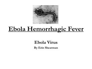
Marburg and Ebola Viruses 1 • Marburg virus was first isolated in 1967 from several cases of hemorrhagic fever in European laboratory workers in Germany and former Yugoslavia working with tissues and blood from African green monkeys imported from Uganda. • Ebola virus was first reported simultaneously in Zaire and Sudan in 1976 when two distinct subtypes were isolated in two hemorrhagic fever epidemics. • Both subtypes, later named Zaire and Sudan, caused severe disease and mortality rates greater than 50%.
Marburg and Ebola Viruses 2 • A third subtype of Ebola (Reston) was later found in macaques imported from the Philippines into the US in 1989 and Italy in 1992. • Four humans were asymptomatically infected and recovered without any signs of hemorrhagic fever. • In 1994, a fourth subtype of Ebola was isolated from an animal worker in Côte d'Ivoire who had preformed a necropsy on an infected chimpanzee. • Scattered outbreaks have occurred periodically with latest being an outbreak of Ebola in the Republic of the Congo in 2007.
Marburg and Ebola Viruses 3 • The reservoir for filoviruses is still unknown. • Bats have been implicated for Marburg virus, but no evidence of Ebola viruses have been found in over 3000 species of animals tested in the areas of human outbreaks. • Intimate person-to-person contact is the main means of transmission of filoviruses in humans.
Marburg and Ebola Viruses 4 • Nosocomial transmission has been a major problem in outbreaks in Africa through the reuse of needles and syringes and exposure to infected tissues, fluids, and hospital materials. • Aerosol transmission has been observed in primates but does not seem to be a major means in humans. • Marburg and Ebola subtypes Sudan, Zaire, and Côte d'Ivoire appear to be found only in Africa and all three Ebola subtypes have only been isolated from human cases in Africa.
Marburg and Ebola Viruses 5 • Filoviruses cause the most severe hemorrhagic fever in humans. • The incubation period for both Marburg and Ebola is generally 4 to 10 days followed by abrupt onset of fever, chills, malaise, and myalgia. • Bleeding from mucosal membranes, venipucture sites and the gastrointestinal tract occurs followed by DIC. • The patient rapidly deteriorates and progresses to multisystem failure. • Death or clinical improvement usually occurs around day 7 to 11.
Marburg and Ebola Viruses 6 • The case fatality rate for Marburg ranges from 23-33% and 53-88% for Ebola with the highest rates found in Ebola Zaire. • Survivors of the hemorrhagic fever are often plagued with arthralgia, uveitis, psychosocial disturbances, and orchitis for weeks following the initial fever. • The presence of Ebola Reston in macaques from the Philippines marked the first time a filovirus was found in Asia. • The pattern of disease of humans in nature is relatively unknown except for major epidemics.
Marburg and Ebola Viruses 7 • Filoviruses cause severe hemorrhagic fever in non-human primates. • The signs and symptoms found are identical to humans. • The only major difference is Ebola Reston has a high mortality in primates (~82%) while it does not seem to be pathogenic to humans.
Yellow Fever Virus 1 • Yellow Fever virus was the first flavivirus to be isolated in 1927 and the first virus to be proved to be transmitted by an arthropod vector. • Yellow Fever was first described in 1648 in Yucatan. • It later caused huge outbreaks in tropical Americas in 17th, 18th, 19th, and 20th century. • The French failed to complete the Panama Canal because their work force was decimated by Yellow Fever.
Yellow Fever Virus 2 • Yellow Fever is a zoonotic diseases that is maintained in non-human primates. • The virus is passed from primate to primate through the bite of the mosquito. • This is known as the sylvatic cycle. • Humans contract the disease when bitten by an infected mosquito usually Aedes aegypti and the disease can then spread from human to human by these mosquitoes. • This cycle is known as the urban cycle.
Yellow Fever Virus 3 • Yellow Fever virus is found throughout sub-Saharan Africa and tropical South America but activity is intermittent and localized. • Yellow Fever is maintained in non-human primates. • The annual incidence is believe to be about 200,000 cases per year globally. • Yellow Fever can cause a severe hemorrhagic fever. The incubation period in humans is 3 to 6 days. • The clinical manifestations can range from mild to severe. Usually, jaundice, proteinuria and hemorrhage
Yellow Fever Virus 4 • Severe Yellow Fever begins abruptly with fever, chills, severe headache, lumbosacral pain, generalized myalgia, anorexia, nausea and vomiting, and minor gingival hemorrhages. • A period of remission may occur for 24 hours followed by an increase in the severity of symptoms. Death usually occurs on day 7 – 10. • Case fatality rate varies greatly depending on the epidemic but may reach up to 50% in severe yellow fever cases.
Dengue Virus 1 • Dengue virus which was also found to be transmitted by an arthropod (Aedes aegypti) was isolated in 1943. • Major outbreaks of dengue with hemorrhagic fever have occurred in Australia in 1897, Greece in 1928, and Formosa 1931. • Since the cessation of the use of DDT to control mosquito vectors, dengue has now spread to most of the tropical regions of the world. • Dengue has been isolated from several non-human primates in Africa but does not cause clinical signs.
Dengue Virus 2 • Dengue virus is found throughout the tropical Americas, Africa, Australia, and Asia. • Fever, muscle and joint pain, lymphadenopathy and rash. • Cases of Dengue Hemorrhagic Fever (DHF) have been increasing as the distribution of Aedes aegypti increases following the collapse of mosquito control efforts. • Case fatality rates for DHF is generally low 1-10% depending on available treatment.
Dengue Virus 3 • Dengue virus will cause a mild flu-like illness upon first exposure. • If the person is then infected by a different serotype, dengue hemorrhagic fever can occur. • The disease will begin like a normal infection of dengue virus with an incubation period of 2-5 days but will quickly progress to a hemorrhagic syndrome. • Rapid shock ensues but can be reversed with appropriate treatment.
Kyasanur Forest Hemorrhagic Fever 1 • Kyasanur Forest virus was isolated from a sick monkey in the Kyasanur Forest in India in 1957. • Since its recognition 400 to 500 cases a year have been reported • Kyasanur Forest virus is transmitted by an ixodid tick. • Livestock may develop viremia with Kyasanur Forest Disease Virus but generally do not show clinical signs. • The basic transmission cycle involves ixodid ticks and wild vertebrates, principally rodents and insect-eating animals.
Kyasanur Forest Hemorrhagic Fever 2 • Kyasanur Forest virus in humans is characterized by fever, headache, myalgia, cough, bradycardia, dehydration, hypotension, gastrointestinal symptoms, and hemorrhages. • Case fatality rate is 3 -5%. • Recovery is generally uncomplicated with no lasting sequelae. • Kyasanur Forest virus is confined to Mysore State of India but spreading.
Omsk Hemorrhagic Fever 1 • Omsk hemorrhagic fever virus was first isolated in 1947 from the blood of a patient with hemorrhagic fever during an epidemic in Omsk and Novosibirsk Oblasts of the former Soviet Union. • The basic transmission cycle of the Omsk Hemorrhagic Fever virus is unknown. • An ixodid tick is believed to transmit the viruses from rodent to rodent. Humans become infected when bitten by an infected tick. • Muskrats are epizootic hosts, and human infections occur by direct contact with their urine, feces, or blood.
Omsk Hemorrhagic Fever 2 • Omsk Hemorrhagic Fever virus is still isolated to the Omsk and Novosibirsk regions of the former Soviet Union. • Omsk Hemorrhagic Fever virus has a similar presentation to Kyasanur Forest virus, however, hearing loss, hair loss, and neuropsychiatric complaints are commonly reported following recovery • Case fatality is 0.5 – 3%. • Omsk Hemorrhagic Fever Virus is maintained in rodents but does not cause clinical signs.