Viral Hemorrhagic Fever
680 likes | 1.28k Vues
Viral Hemorrhagic Fever. Overview. Organism History Epidemiology Transmission Disease in Humans Disease in Animals Prevention and Control. What is Viral Hemorrhagic Fever?. Severe multisystem syndrome Diffuse Damage to overall vascular system

Viral Hemorrhagic Fever
E N D
Presentation Transcript
Overview • Organism • History • Epidemiology • Transmission • Disease in Humans • Disease in Animals • Prevention and Control
What is Viral Hemorrhagic Fever? • Severe multisystem syndrome • Diffuse Damage to overall vascular system • Symptoms often accompanied by hemorrhage • Rarely life threatening in itself • Includes conjunctivitis, petechia, echymosis • Relatively high mortality
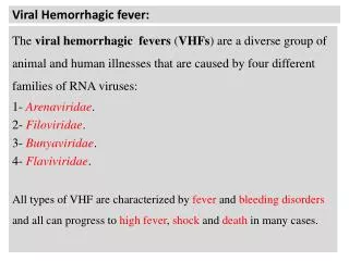
Quick Overview: Who are they? • VHFs are: • Enveloped Lipid-encapsulated • Single-strand RNA • Zoonotic (animal-borne) • Geographically restricted by host • Persistent in nature (rodents, bats, mosquitoes, ticks, livestock, monkeys, and primates) • Survival dependent on an animal or insect host, for the natural reservoir
Arenaviridae Lassa Fever Argentine HF (Junin) Bolivian HF (Machupo) Brazilian HF (Sabia) Venezuelan HF (Guanarito) Bunyaviridae Rift Valley Fever (RVF) Crimean Congo HF (CCHF) Hantavirus (Hemorrhagic Fever with Renal Syndrome (HFRS)) Hantavirus Pulmonary Syndrome (HPS) Filoviridae Marburg Ebola Flaviviridae Yellow Fever Dengue Fever Omsk HF Kyasanur Forest Disease Quick Overview: Who are they?
Quick Overview: How do we get infected? • Rodents & Arthropods, both reservoir & vector • Bites of infected mosquito or tick • Inhalation of rodent excreta • Infected animal product exposure • Person-to-Person • Blood/body fluid exposure • Airborne potential for some arenaviridae, filoviridae
Arenaviridae Junin virus Machupo virus Guanarito virus Lassa virus Sabia virus
Arenaviridae History • First isolated in 1933 • 1958: Junin virus - Argentina • First to cause hemorrhagic fever • Argentine hemorrhagic fever • 1963: Machupo virus – Bolivia • Bolivian hemorrhagic fever • Guanarito (Venezuela) • Sabia (Brazil) • 1969: Lassa virus – Nigeria • Lassa fever
Arenavirus Structure • Single-stranded, bi-segmented RNA genome • Large segment (7200nt), small one (3500nt) • Lipid envelope with 8-10nm club-shaped projections
Arenaviridae Transmission • Virus transmission and amplification occurs in rodents • Shed virus through urine, feces, and other excreta • Human infection • Contact with excreta • Contaminated materials • Aerosol transmission • Person-to-person transmission
Arenaviridae in Humans • Incubation period 10–14 days • Fever and malaise 2–4 days • Hemorrhagic stage • Hemorrhage, leukopenia, thrombocytopenia • Neurologic signs
Arenaviridae: Lassa Fever • First seen in Lassa, Nigeria in 1969. • Now in all countries of West Africa • 5-14% of all hospitalized febrile illness • Rodent-borne (Mastomysnatalensis) • Interpersonal transmission • Direct Contact • Sex • Breast Feeding
Lassa Fever Virus • Background • Discovered in 1969 when two missionary nurses died in Lassa, Nigeria, W. Africa • It expands to Guinea, Liberia, Sierra Leone • 100 to 300 thousand cases per year with approx. 5,000 deaths
Distinguishing Features Gradual onset Retro-sternal pain Exudative pharyngitis Hearing loss in 25% may be persistent Spontaneous abortion Mortality 1-3% overall (up to 50% in epidemics) Therapy: Ribavirin Lassa Fever
Bunyaviridae L-segment codes for an L-protein (the RNA dependent RNA polymerase); M segment codes for two surface glycoproteins G1 and G2 which form the envelope spikes; S segment codes for an N-protein (nucleocapsid protein). Rift Valley Fever virus Crimean-Congo Hemorrhagic Fever virus Hantavirus
Bunyaviridae • Rift Valley Fever (RVF) • Crimean-Congo Hemorrhagic Fever (CCHF) • Hantavirus • Old World: Hemorrhagic fever with renal syndrome (HFRS) • New World: Hantavirus pulmonary syndrome (HPS) • 5 genera with over 350 viruses
Bunyaviridae Transmission • Arthropod vector • Exception – Hantaviruses • RVF – Aedes mosquito • CCHF – Ixodid tick • Hantavirus – Rodents • Less common • Aerosol • Exposure to infected animal tissue
Bunyaviridae • Transmission to humans • Arthropod vector (RVF, CCHF) • Contact with animal blood or products of infected livestock • Rodents (Hantavirus) • Laboratory aerosol • Person-to-person transmission with CCHF
Rift Valley Fever • Predominantly a disease of sheep and cattle • 1930: First identified in an infected newborn lamb in Egypt • In livestock: • ~100% abortion • 90% mortality in young • 5-60% mortality in adults
Asymptomatic or mild illness in humans Distinguishing Characteristics Hemorrhagic complications rare (<5%) Vision loss (retinal hemorrhage, vasculitis) in 1-10% Overall mortality 1% Therapy: Ribavirin? Rift Valley Fever
Crimean-Congo Hemorrhagic Fever • Distinguishing features • Abrupt onset • Most humans infected will develop hemorrhagic fever • Profuse hemorrhage • Mortality 15-40% • Therapy: Ribavirin
Bunyaviridae: Crimean-Congo HF • Transmission to humans: • Ixodid, Hyalomma spp. ticks • Contact with animal blood/products • Person-to-person • Laboratory aerosols • Extensive geographical distribution
Bunyaviridae: Hantaviruses • Transmission to humans: • Exposure to rodent saliva and excreta • Inhalation • Bites • Ingestion in contaminated food/water (?) • Person-to-person (Andes virus in Argentina)
Hemorrhagic Fever with Renal Syndrome (HFRS) • Distinguishing Features • Insidious onset • Intense headaches, • Blurred vision • kidney failure • (causing severe fluid overload) • Mortality: 1-15%
Bunyaviridae Humans • RVF • Incubation period – 2-5 days • 0.5% - Hemorrhagic Fever • CCHF • Incubation period – 3-7 days • Hemorrhagic Fever - 3–6 days following clinical signs • Hantavirus • Incubation period – 7–21 days • HPS and HFRS
Filoviridae • Ebola • Ebola-Zaire • Ebola-Sudan • Ebola-Ivory Coast • Ebola-Bundibugyo • (Ebola-Reston) • Marburg Ebola Marburg
Filoviridae History • 1967: Marburg, Frankfurt, Belgrade • European laboratory workers • 1976: Ebola virus • Ebola Zaire • Ebola Sudan • 1989 and 1992: Ebola Reston • USA and Italy • Imported macaques from Philippines • 1994: Ebola Côte d'Ivoire
Filoviridae Transmission • Reservoir is UNKNOWN • Bats implicated with Marburg • Intimate contact • Nosicomial transmission • Reuse of needles and syringes • Exposure to infectious tissues, excretions, and hospital wastes • Aerosol transmission • Primates
Filoviridae: Ebola • Rapidly fatal febrile hemorrhagic illness • Transmission: • bats implicated as reservoir • Person-to-person • Nosocomial • Five subtypes • Ebola-Zaire, Ebola-Sudan, Ebola-Ivory Coast, Ebola-Bundibugyo, Ebola-Reston • Ebola-Reston imported to US, but only causes illness in non-human primates • Human-infectious subtypes found only in Africa
Distinguishing features: Acute onset Weight loss/protration 25-90% case-fatality Filoviridae: Ebola
Filoviridae: Marburg • Transmission: • Animal host unknown • Person-to-person • infected animal blood/fluid exposure • Indigenous to Africa • Uganda • Western Kenya • Zimbabwe • Democratic Republic of Congo • Angola
Distinguising features Sudden onset Chest pain Maculopapular rash on trunk Pancreatitis Jaundice 21-90% mortality Filoviridae: Marburg
Filoviridae Humans • Most severe hemorrhagic fever • Incubation period: 4–10 days • Abrupt onset • Fever, chills, malaise, and myalgia • Hemorrhage and DIC • Death around day 7–11 • Painful recovery
Flaviviridae Dengue virus Yellow Fever virus Omsk Hemorrhagic Fever virus Kyassnur Forest Disease virus
Flaviviridae History • 1648 : Yellow Fever described • 17th–20th century • Yellow Fever and Dengue outbreaks • 1927: Yellow Fever virus isolated • 1943: Dengue virus isolated • 1947 Omsk Hemorrhagic Fever virus isolated • 1957: Kyasanur Forest virus isolated
Flaviviridae Transmission • Arthropod vector • Yellow Fever and Dengue viruses • Aedesaegypti • Sylvatic cycle • Urban cycle • Kasanur Forest Virus • Ixodid tick • Omsk Hemorrhagic Fever virus • Muskrat urine, feces, or blood
Flaviviridae Epidemiology • Yellow Fever Virus – Africa and Americas • Case fatality rate – varies • Dengue Virus – Asia, Africa, Australia, and Americas • Case fatality rate – 1-10% • Kyasanur Forest virus – India • Case fatality rate – 3–5% • Omsk Hemorrhagic Fever virus – Europe • Case fatlity rate – 0.5–3%
Flaviviridae Humans • Yellow Fever • Incubation period – 3–6 days Short remission • Dengue Hemorrhagic Fever • Incubation period – 2–5 days • Infection with different serotype • Kyasanur Forest Disease • Omsk Hemorrhagic Fever Lasting sequela
Yellow Fever • Distinguishing features • Biphasic infection • Common hepatic involvement & jaundice • Mortality: 15-50%
Flaviviridae: Dengue • Dengue Fever (DF) /Fatality:<1% • Dengue Hemorrhagic Fever (DHF)/ Fatality: 5-6% • Dengue Shock Syndrome (DSS)/Fatality 12-44% • Four distinct serotypes • DEN-1, DEN-2, DEN-3, DEN-4 • Distinguishing Features • Sudden onset • Eye pain • Rash • Complications/sequelae uncommon • Illness less severe in younger children
Omsk Hemorrhagic Fever • Distinguishing Features • Acute Onset • Biphasic infection • Complications • Hearing loss • Hair loss • Psycho-behavioral difficulties • Mortality: 0.5 – 3%
Distribution: limited to Karnataka State, India Distinguishing Features Acute onset Biphasic Case-fatality: 3-5% (400-500 cases annually) Flaviviridae: Kyanasur Forest
Common Pathophysiology • Small vessel involvement • Increased vascular permeability • Multiple cytokine activation • Cellular damage • Abnormal vascular regulation: • Early -> mild hypotension • Severe/Advanced -> Shock • Viremia • Macrophage involvement • Inadequate/delayed immune response
Common Pathophysiology • Multisystem Involvement • Hematopoietic • Neurologic • Pulmonary • Hepatic (Ebola, Marburg, RVF, CCHF, Yellow Fever) • Renal (Hantavirus) • Hemorrhagic complications • Hepatic damage • Consumptive coagulopathy • Primary marrow injury to megakaryocytes
Fever Myalgia Malaise Fatigue/weakness Headache Dizziness Arthralgia Nausea Non-bloody diarrhea Common Clinical Features: Early/Prodromal Symptoms
Conjunctivitis Facial & thoracic flushing Pharyngitis Exanthems Periorbital edema Pulmonary edema Hemorrhage Subconjunctival hemorrhage Ecchymosis Petechiae But the hemorrhage itself is rarely life-threatening. Common Clinical Features: Progressive Signs
Symptoms • Incubation period of 6-21 days • 80% of human infections are asyptomatic • Onset is slow: fever, weakness, & malaise • Few days: headache, pharyngitis, muscle pain, retrostinal & abdominal pain, nausea, vomiting, conjunctivitis, diarrhea, cough, & proteinuria • Severe cases: • facial swelling, lung cavity fluid, hemorrhaging, hyopotension, • Neurological problems: tremors, encephalitis, hair loss, gait disturbance, deafness • 95% death rate among pregnant women & spontaneous abortion
Multisystem compromise Profuse bleeding Consumptive coagulopathy/DIC Encephalopathy Shock Death Common Clinical Features: Severe/End-stage
Clinical Symptoms • More severe • Bleeding under skin • Petechiae, echymoses, conjunctivitis • Bleeding in internal organs • Bleeding from orifices • Blood loss rarely cause of death