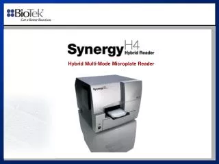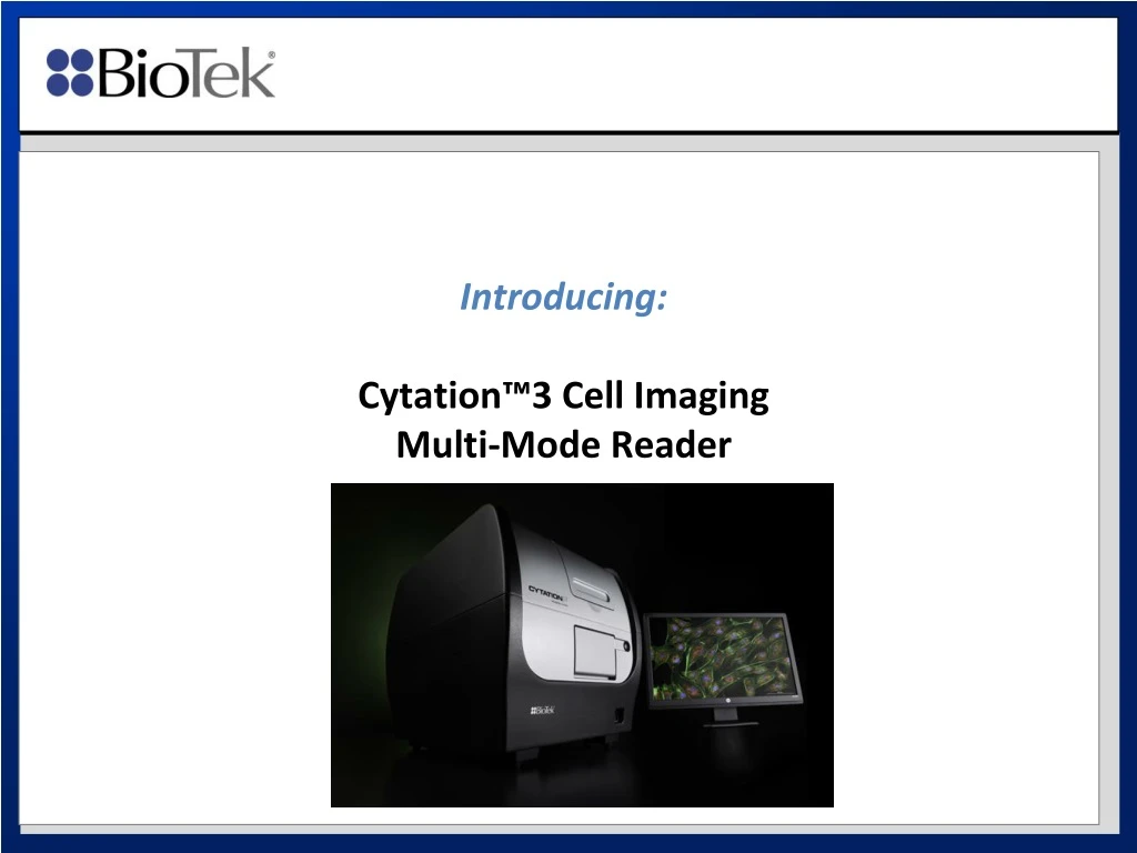
Introducing: Cytation™3 Cell Imaging Multi-Mode Reader
370 likes | 417 Vues
Introducing Cytation™3, an affordable and easy-to-use microplate imager that supports increased cell biology research with quantitative and qualitative data. With live cell features and advanced image analysis capabilities, Cytation™3 provides a fuller picture for cell biology research.

Introducing: Cytation™3 Cell Imaging Multi-Mode Reader
E N D
Presentation Transcript
Project Goals • Main project goal was to design: • An affordable microplate imager to support increased cell biology research • A combined microplate reader / imager that provides new benefits such as a unique hit-picking feature • Easy-to-use software interface to reduce learning curve for non-imaging experts
Traditional Plate Reader - Filters • Quantitative data and sensitivity • Precisely measure cellular activity
Traditional Plate Reader - Monochromator • Quantitative data and flexibility • Precisely measure cellular activity
Imaging Optical Path • Qualitative and semi-quantitative data • Visualize change in phenotype
Microplate Reader with Imaging • Quantitative numerical data • Quantitative numerical data • Fuller picture for cell biology research • Qualitative phenotypic data • Qualitative data
More and more assays are run on live cells • Cytation3 live cell features: • Temperature control up to 45ºC • Gas controller (CO2 / O2) • Automatic reagent dispenser • Quantitative numerical data • Qualitative phenotypic data
Fluorescence Optical Path - Widefield 4. Samples 5. Gray scale CCD camera 3. Objective CCD 2. Filter/mirror cube 1. Light source (LED)
Bright Field Optical Path 1. Light source (LED) 2. Samples CCD 4. Gray scale CCD camera 3. Objective
8 Colors Available Right Away DAPI GFP Texas Red RFP YFP CY5 CY7 CFP Custom…
BioTek objectives (also called “lenses”): Zeiss 2.5 x Olympus 4 x Olympus 10 x Olympus 20 x No-brand 2x
Magnification vs. well size 2.5x 4x 10x 20x 2x 96-well 6.5 mm 384-well 3.7 mm
Magnification vs. pixel size 2.5x 3 x 2.3 mm 4x 2 x 1.5 mm 10x 0.8 x 0.6 mm 20x 0.4 x 0.3 mm 384-well 3.7 mm
200 x What does 2.5x, 4x, 10x… mean? Objective magnification 50 x Computer monitor (example: 40 x 30 cm) 4 x CCD sensor (about 8 x 6 mm) Measurement area in the well (2.0 x 1.5 mm) 200 x 800 x Projection screen (160 x 120 cm)
Numerical aperture (NA) and Working Distance Microplate applications typically require long working distance objectives
Magnification and example of applications 20x 0.4 x 0.3 mm 4x 2 x 1.5 mm 2.5x 3 x 2.3 mm • No cell details at all • Counting could be difficult (many overlapping objects) • In this case, total image intensity would be used rather than count. • Limited cell details • Allows counting, and many cells are visible so good statistics • Cytotoxicty, cell proliferation assays • Intracellular details visible • Qualitative information about location of fluorescence in cells • Advanced image analysis can be performed in Gen5 or third party software. • Apoptosis, translocation events…
Only One Firewire Port is Used (and Visible on the Instrument)
Firewire Port Card (Laptop or Desktop) Why FireWire? High Data Transfer Speed. What about smaller laptop Firewire Ports? No Power.
CCD chip and Pixels CCD sensor / chip Sony chip Number of pixels: 1384 x 1036 (sensor) 1280 x 960 (image) Pixel size 4.65 um Each image contains about 1,2 million pixels
“Bit depth” Pixel value = 0 (black) Pixel value = 22,645 (gray) Pixel value = 65,535 (white) • Common camera bit depth: • 8-bit = 255 values (0=black, 255 = white) • 12-bit = 4095 values (0=black, 4095 = white) • 16-bit = 65535 values (0=black, 65535 = white)
Gray scale • No color information is stored per pixel: gray scale image • Colors are added after the fact by Gen5 (see next slide) • A pixel at 65535 is considered “saturated” (equivalent to “overflow” on PMT), but pixel value will still be set to 65535 (software will have ability to identify and highlight saturated pixels).