Download
1 / 74
770 likes | 1.25k Vues
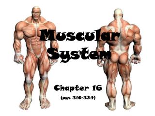
Muscular System. http://www.youtube.com/watch?v=Ry9IcQMFiz8&feature=fvw. How many muscles do we have?. Over 650!. Our over 650 muscles fall into three categories – 1. skeletal, 2. cardiac & 3. smooth. Fun Facts. There are 50 muscles in your face alone.

E N D
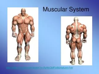
Muscular System http://www.youtube.com/watch?v=Ry9IcQMFiz8&feature=fvw
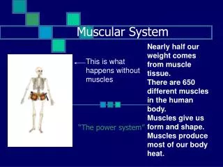
How many muscles do we have? Over 650! • Our over 650 muscles fall into three categories – • 1. skeletal, • 2. cardiac & • 3. smooth.
Fun Facts • There are 50 muscles in your face alone. • It takes 17 muscles to smile and 40 muscles to frown. • Your heart muscle NEVER gets any rest neither do the muscles of your intestines! • The most active muscles are those in your eyes. They move more than 100,000 times a day!
Biggest, Strongest, and Hardest Working • The gluteus maximusis the largest muscle in the human body. • The strongest muscle in your body is the masseter, located on each side of your mouth. These muscles help you bite down with 150 pounds of force! • The hardest working muscle is the heart. Every day the heart pumps at least 2,500 gallons of blood.
Three Types of Muscle Cells • Skeletal • Cardiac • Smooth
Skeletal Muscle a.k.a. striated muscle Nuclei are on the periphery of the cells
3. What kind of muscle? Do Now! 1. What kind of muscle? (C, Sm, Sk?) (C, Sm, Sk?) 4. Lines hollow organs (C, Sm, Sk?) 5. Makes up the heart walls (C, Sm, Sk?) 6. Connected to bone (C, Sm, Sk?) 7. Involuntary (C, Sm, Sk?) 8. Used to push substances along internal passageways. (C, Sm, Sk?) 9. Voluntary (C, Sm, Sk?) 10. Moves bones (C, Sm, Sk?) 2. What kind of muscle? (C, Sm, Sk?)
Practice Practical Muscle Quiz • 1. Identify the type of tissue. • A) Dense regular connective tissue • B) Smooth muscle • C) Cardiac muscle • D) Skeletal muscle • 2. In question 1, above, what was the most distinctive identifying feature? • Striations • Branched fibers • Peripheral nuclei • A and C • A, B, and C
3. Identify this tissue: A) Dense regular connective tissue B) Smooth muscle C) Cardiac muscle D) Skeletal muscle 4. In question 3, above, what was the most distinctive identifying feature? A) Striations B) Branched fibers C) Central nuclei D) A and C E) A, B, and C
5. This specialized junction separates cells in: A) Cardiac muscle B) Skeletal muscle C) Smooth muscle D) Tendons
6. Identify this tissue. A) Dense regular connective tissue B) Smooth muscle C) Cardiac muscle D) Skeletal muscle E) Dense irregular connective tissue
7. Identify this tissue. A) Dense regular connective tissue B) Smooth muscle C) Cardiac muscle D) Skeletal muscle E) Dense irregular connective tissue
8. Identify this tissue: A) Dense regular connective tissue B) Smooth muscle C) Cardiac muscle D) Skeletal muscle E) Dense irregular connective tissue 9. In question 8, above, what was the most distinctive identifying feature? A) Striations B) Branched fibers C) Peripheral nuclei D) A and C E) A, B, and C
10. Identify the name of the connective tissue around each fiber: A) epimysium B) endomysium C) perimysium D) meromysium E) sarcomysium
11. Identify the structures noted by the arrows: A) striations B) sarcomeres C) Intercalated discs D) Sarcoplasmic reticulum E) endomysium
Skeletal Muscle Fibers • Connect to bones • Forms the smoother contours of the body • Very long and cylindrical in shape • Multinucleated • Largest of the 3 muscle types • A.K.A. Striated muscles • Only muscle type under voluntary control • Can also be activated by automatic reflexes
How can they be so strong? • Each fiber is wrapped in connective tissue called endomysium. • Several fibers are then wrapped in a courser membrane called a perimysium to form a fascicle. • The fascicle is bound together by an even tougher cover called an epimysium. Endomysium
The epimysium (outer layer) can blend into strong cordlike tendonsor • Into sheet-like aponeuroses (fascia) which attach muscle indirectly to bone, cartilages, or other connective tissue coverings.
Label this skeletal muscle using the diagram on the next slide
Origin vs. Insertion • The muscles origin is attached to the immovable bone. • At its other end, the insertion is attached to the movable bone. You’ll need this for lab!
The myofilaments of a myofibril are arranged in a regular fashion so that their ends are all lined up. This is what gives the muscle its striated appearance. The contractile units of the cells are called sarcomeres.
The myofibrils have distinct, repeating microanatomical units, termed sarcomeres. Sarcomere The sarcomere is composed of thick and thin filaments – myosin and actin, respectively. Chemical and physical interactions between the actin and myosin cause the sarcomere length to shorten (contract). The interactions between actin and myosin serve as the basis for the sliding filament theory of muscle contraction.
Sliding Filament Theory • The sliding filament theory is the basic summary of the process of skeletal muscle contraction. • Myosin moves along the filament by repeating a binding and releasing sequence that causes the thick filament to move over the thinner filament. • This progresses in sequential stages. By progressing through this sequence the filaments slide and the skeletal muscles contract and release.
Naming Skeletal Muscles • Named based on structural or functional characteristics. 1. Direction of fibers: Rectus (straight or parallel to an imaginary line) or oblique (at a slant)
Naming Skeletal Muscles 2. Size of muscle: Maximus (largest), Minimus (smallest) and Longus (long)
Naming Skeletal Muscles 3. Location of the Muscle Some are named for the bone they attach are associated with. Ex. The temporalis muscle lays over the temporal bones of the skull.
Naming Skeletal Muscles 4. Number of Origins Ex. Bicep muscle has two heads, triceps have three and quadriceps have four.
Naming Skeletal Muscles 5. Location of Muscle’s origin and insertion. • Ex. Sternocleidomastoid has its origin on the sternum (sterno), and clavicle (cleido) and inserts on the mastoid process of the temporal bone.
Naming Skeletal Muscles 6. Shape of the Muscle If a muscle has a definitive shape, it is used to help identify them. Ex. Deltoid (triangular)
Naming Skeletal Muscles 7. Action of the Muscle Adductor: A motion that pulls a structure or part towards the midline of the body, or towards the midline of a limb Flexor: Closes a joint Bending movement that decreases the angle between two parts. For ex. bending the elbow, or clenching a hand into a fist Extensor: opens a joint a straightening movement that increases the angle between body parts Abductor: moves away from midline
Four functions of skeletal muscles. • Maintain posture • Stabilize joints • Generate heat • Produce movement
Aging and Muscles • After 30, your muscle mass dwindles some 3-8 percent each decade. Once you hit 60, these losses accelerate even more quickly. • Decreased muscle mass means you'll burn far fewer calories. • Muscles require a lot of calories to maintain. (Think of them as a bunch of high-strung, active family members visiting your home. They're always up, moving around. As a result, they're hungry and require a lot of food.) • The strength of your muscles is related to the strength of your bones. When your muscles are weak, your bones are more likely to be weak. (This is esp. important for women who have higher risk of osteoporosis.)
Muscle Pairs • Muscles are usually in (Antagonistic) pairs. When one muscle contracts, the other extends. • Adrenaline allows your muscles to use 4 to 6 times more oxygen than usual thus creating a huge amount of energy. • Extreme strength:http://www.youtube.com/watch?v=Q9wRTIZIByk&NR=1 • Two man strength performance:http://www.youtube.com/watch?v=JWK5mfRGkiE&feature=related
Disorders of the Muscular System • Muscular Dystrophy: Occurs when a particular gene on the X chromosome: a. fails to make the protein dystrophin. b. makes low amounts of dystrophin. • the membranes around muscle cells become weak and tear easily, eventually leading to the death of muscle fibers. • More common in males.
DMD Types of MD A. When one of these proteins, dystrophin, is absent, the result is Duchene’s muscular dystrophy. B. Poor or inadequate dystrophin results in Becker’s muscular dystrophy. Animation: http://www.youtube.com/watch?v=6wLnR7GJakY A family’s story: http://www.youtube.com/watch?v=dSEc5zvGTbc
Disorders of the Muscular System 2. ALS: Amyotrophic lateral sclerosis: disease of the nerve cells in the brain and spinal cord that control voluntary muscle movement. • AKA Lou Gehrig's disease. • The symptoms usually do not develop until after age fifty. • Persons with this disease have a loss of muscle strength and coordination that eventually gets worse. • Breathing or swallowing muscles may be the first muscles affected. As the disease gets worse, more muscle groups develop problems. • Life expectancy of an ALS patient averages about two to five years from the time of diagnosis.
Amyotrophic lateral sclerosis (ALS) is caused by the degeneration and death of motor neurons in the spinal cord and brain. These neurons convey electrical messages from the brain to the muscles to stimulate movement in the arms, legs, trunk, neck, and head. As motor neurons degenerate, the muscles are weakened and cannot move as effectively, leading to muscle wasting.
http://www.youtube.com/watch?v=tKRWUZr4gBg Fans, for the past two weeks you have been reading about the bad break. Today I consider myself the luckiest man on the face of this earth. That I might have been given a bad break, but I have an awful lot to live for. Thank you. His record for most career grand slam home runs (23) still stands today (Alex Rodriguez has 22, Manny Ramirez has 21)
Disorders of the Muscular System 3. Myasthenia gravis: is caused by a defect in the transmission of nerve impulses to muscles. • Its a disease in which the immune system attacks the body's own tissues ("autoimmune" disease); • Causes a weakness of voluntary muscles without pain that gets worse with repeated or sustained use of the muscle (fatigued muscle weakness). • In two thirds of patients with MG, the first muscles to be affected are those controlling eye movements (causing double vision) and those holding the eyelids up (causing drooping of the eyelids). • There are medications to treat the disorder. It is not fatal.
4. Chronic Fatigue Syndrome: Symptoms of chronic fatigue syndrome include loss of memory, difficulty concentrating, fatigue, random muscle pain, headaches, unrefreshing sleep and sore throats. 5. Fibromyalgia: results in widespread pain throughout every muscle in a person's body. Approximately 2% of the entire US population is affected by fibromyalgia. Symptoms of fibromyalgia include joint tenderness, fatigue problems, and sleep disturbances. No cure.
6. Cerebral Palsy: • Cerebral palsy is condition that can involve brain and nervous system functions such as movement, learning, hearing, seeing, and thinking. • There are several different types of cerebral palsy. • Cerebral palsy is caused by injuries or abnormalities of the brain. • Most of these problems occur as the baby grows in the womb, but they can happen at any time during the first 2 years of life, while the baby's brain is still developing.
Difficulty swallowing or speaking • Facial weakness on both sides of the face • Blurred vision • Drooping eyelids • Trouble breathing • Nausea, vomiting and abdominal cramps (only in food-borne botulism) • Paralysis Botulism & Botox • Botulism is a rare but serious condition caused by toxins from bacteria called Clostridium botulinum. • can be fatal • A weakened botulinum toxin (BOTOX) has been used to reduce facial wrinkles by paralyzing muscles beneath the skin, and for medical conditions, such as eyelid spasms and severe underarm sweating.