Download
1 / 34
380 likes | 1.1k Vues
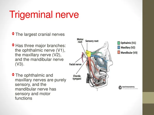
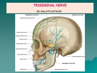
CLINICAL EVALUATION OF TRIGEMINAL NERVE FUNCTION. Sensory Evaluation. Exteroceptive sensation (pain, light touch, heat, and cold) three Vth divisions : tested individually and compared with opposite side .

E N D
Sensory Evaluation • Exteroceptivesensation (pain, light touch, heat, and cold) • three Vthdivisions : tested individually and compared with opposite side. • Lesions of individual divisions (distal to the gasserian ganglion) : result in sensory loss confined to the cutaneous supply of that division • Lesions at or proximal to the gasserianganglion :affects whole I/L face.
Lesions within the brainstem or upper cervical cord :onion-skin distribution of sensory loss, • Dissociation of sensation on the face (pain and temperature vs. touch sensation) differentiates lesions affecting the spinal tract and nucleus of the Vth nerve from lesions affecting the main sensory nucleus. • The cutaneous area over the angle of the mandible is supplied by the second and third cervical roots (by way of the great auricular nerve) and not by the Vth nerve
Motor Evaluation supplies the muscles of mastication. clench the jaw (Ms and T), move the jaw from side to side against resistance (lateral pterygoids), and protrude the jaw.
With nuclear or infranuclear lesions of the motor division of the Vth nerve (LMN ) • I/L T and M muscles do not contract when the jaw is clenched, • the jaw deviates to the paralyzed side when the mouth is opened (due to contraction of the C/L intact LP muscle), and • the jaw cannot be deviated toward the nonparalyzed side (due to I/L LP paresis). • Atrophy and fasciculation of the masticatory muscles may also be evident.
Other muscles supplied by the Vth nerve (mylohyoid, anterior belly of the digastric, tensor tympani, tensor velipalatini) are difficult to evaluate clinically. • Flaccidity of floor of mouth due to mylohyoid and digastric paralysis may be evident on palpation, and • Paralysis of tensor tympani: result in difficulty in hearing high notes.
Reflex Evaluation : • corneal reflex • Sternutatory reflex and • jaw jerk (M reflex).
Corneal reflex: • Afferent arc : travels through the ophthalmic (upper cornea) and maxillary (lower cornea) • Efferent arc : I/L(direct reflex) and C/L (consensual reflex) facial nerve to the orbicularisoculi muscles. • Lesions of Vth nerve (peripheral or pontomedullary trigeminal pathways) result in loss of I/L and C/L responses. • Suprasegmental modulation of this reflex : a parietal lobe lesion (involving the perisylvian portion of the postcentralgyrus) may result in a C/L loss of the corneal reflex
Sternutatory reflex • Afferent : VI • Efferent : v , vii , ix, x, xi and motor nerves of cervical and thoracic spinal cord • Center – brainstem and upper spinal cord • Use : cross check on corneal reflex • Photo-sternutatory / photic sneeze ( ACHOO – AD compelling helio-ophthalmic outburst ) : sneezing on looking at bright light .
Jaw jerk • Involves contraction of the M and T muscles when the patient's lower jaw is tapped. • Afferent arc :1a motor fibers in V2 division • Center :mesencephalic nucleus of the Vth nerve. • Efferent arc :also travels through mandibularfibers that originate in the motor nucleus of the Vth nerve. • Lesions anywhere along this reflex arc • depression of the I/L jaw reflex, • whereas B/L supranuclear lesions result in accentuated response.
Blink reflex • Blink reflex (glabellar reflex, orbicularisoculi reflex): • Percussion over the supraorbital ridge results in B/L contraction of the orbicularisoculi muscles. • afferent arc -mediated by I/L main sensory nucleus of the Vth nerve and the I/L and C/L spinal nuclei of the Vth nerve. • The spinal nuclei of the Vth nerve (B/L) make motor connections through the corresponding facial nuclei, which innervate the orbicularisoculi muscles. • By studying the blink reflex electrically, subtle peripheral and central lesions of the Vth and facial nerves may be uncovered.
Corneo-mandibular reflex • Consists of B/L eye blink and a brisk C/L anterolateral jaw movement induced by corneal stimulation • ? Associated movt rather than true reflex • s/o supranuclear interruption of I/L cortico-trigeminal tract ( only eye sign in ALS ) • In the corneomandibular reflex, the jaw movement is primarily related to the blink rather than the corneal stimulus
Localization of Lesions Affecting Cranial Nerve V SupranuclearLesions • Supranuclear control of Vthmotor function is B/L; however, the C/L hemisphere exerts predominant control on the voluntary activity of the M • Corticobulbarfibers originate in the lower frontal motor cortex, descend through the CR, IC, and cerebral peduncle, and then decussate in the ponsto supply the motor nucleus of the Vthnerve. • Lesions interrupting this pathway may result in C/L Vthmotor paresis (e.g., deviation of the jaw away from the lesion), but because of the B/L innervation, paresis may be mild.
Supranuclear Lesion : • B/L upper motor neuron lesions (pseudobulbar palsy) result in profound Vth motor paresis, often with an exaggerated jaw reflex. Mastication is then markedly impaired. • Thalamic lesions may result in anesthesia of the C/L face. • Parietal lesions may be a/w depression of the C/L corneal reflex, even when facial sensation is otherwise intact
Nuclear Lesions • Involve other brainstem structures, and therefore • long tract signs, and • other cranial nerve involvement • I/L paresis, atrophy, and fasciculations of the muscles of mastication occur.
A pontine localization of this masticatory paresis is suggested by associated findings: • contralateralhemiplegia (due to affection of the basis pontis), • ipsilateralhemianesthesia of the face (due to affection of the main sensory nucleus of the trigeminal nerve), • contralateralhemisensory loss of the limbs and trunk (due to spinothalamic tract affection), and • Internuclearophthalmoplegia (secondary to medial longitudinal fasciculus damage) and • ipsilateral Horner syndrome (due to involvement of descending sympathetic fibers)
The nucleus of the spinal tract of the trigeminal nerve extends from the caudal end of the pons to the third or fourth cervical spinal cord level. • Therefore, lesions affecting the caudal pons, lateral medulla, or upper cervical cord result in : • ipsilateral facial analgesia, hypesthesia, and thermoanesthesia. • Because the lateral spinothalamic tract lies in close proximity to the trigeminal spinal nucleus,it is often associated with contralateral trunk and extremity hypalgesia and thermoanesthesia.
onion-skin pattern of sensory loss • Lower medullary or upper cervical spinal nuclear lesions result in sensory disturbance that affects the peripheral (lateral) forehead, cheek, and jaw (onion-skin pattern of sensory loss). • This onion-skin segmental distribution reflects the rostral-caudal somatotopic arrangement of the cutaneous distribution of the spinal nucleus (e.g., perioral area -rostral; lateral face -caudal).
lateral medullary (Wallenberg) syndrome/PICA Syndrome ) • spinal nucleus of the trigeminal nerve is characteristically affected • most often secondary to brainstem infarction due to intracranial vertebral artery occlusion • Classically attributed to PICA occlusion, but in 80-85% of cases the vertebral artery is also involved“. • Acute onset
Lesions affecting the mesencephalic nucleus of the trigeminal nerve cause no apparent neurologic signs and symptoms, except perhaps depression of the ipsilateral jaw jerk (masseter reflex).
Lesions Affecting the Preganglionic Trigeminal Nerve Roots • In its cisternal course, the preganglionic trigeminal nerve root may be damaged by • tumor (meningioma, schwannoma, metastasis, nasopharyngeal carcinoma), • infection (granulomatous, infectious, or carcinomatous meningitis), • trauma, or • aneurysm. • Preganglionic trigeminal nerve involvement is suggested by the involvement of the neighboring cranial nerves (especially cranial nerves VI, VII, and VIII).
The trigeminal roots may be involved by extension of pathologic processes (usually acoustic neuroma or meningioma) located in the cerebellopontine angle. • associated with ipsilateral tinnitus, deafness, and vertigo (due to involvement of cranial nerve VIII). • Facial nerve paralysis, • ipsilateral ataxia, and nystagmus (due to involvement of the cerebellar peduncles and cerebellum), • ipsilateral lateral rectus paralysis (Vith CN involvement), • and, rarely, affection of cranial nerves IX through XII may also occur.
Trigeminal neuralgia (tic douloureux, Fothergill's disease) • syndrome of sudden, excruciating, lancinating, paroxysmal, and usually unilateral pains in the distribution of one or more of the divisions (often the maxillary or mandibular) of the trigeminal nerve • Incidence:4-5/100,000 • Female predominance (m: f = 1:2 ~2:3) • Mean age: 50 yrs. • Right side more than the left. • B/L : exceedingly rare, except in Multiple sclerosis. • Pain from (V2):referred to upper lip, nose, and cheek. • (V3): lower lip. • Tic pain confined to the ophthalmic division (V1) is distinctly uncommon.
Characteristics of trigeminal neuralgia • paroxysms of severe, lancinating, electric shock-like bouts of pain restricted to the distribution of the trigeminal nerve • Unilaterally (right side) • The mandibular (V3) and/or maxillary (V2) branch or, rarely, the ophthalmic (V1) branch • Spontaneous attack or triggered by trigger zone & movement of the face • Seconds to minutes
Triggers may be shaving, brushing teeth, drinking, eating or even slight breeze
Diagnostic criteria for classic trigeminal neuralgia • Paroxysmal attacks of pain lasting from a fraction of a second to two minutes that affect one or more divisions of the trigeminal nerve • Pain has at least one of the following characteristics • intense, sharp, superficial, or stabbing • precipitated from trigger areas or by trigger factors • Attacks are similar in individual patients • No neurological deficit is clinically evident • Not attributed to another disorder
Etiology • Idiopathic (vascular) • Tumors (V nerve schwannoma, CPA tumors) • Human herpes simplex virus • Multiple Sclerosis • 4% patients with MS have TN • 2% patients with TN have MS
Vascular • An aberrant loop of artery, or less commonly vein, is found to be compressing the root entry zone of the trigeminal nerve in 80-90% of patients at surgery • The nerve is demyelinated next to the compressing vessel • Eliminating the compression provides long term relief • Sensory function recovers after decompression • Compression by tumours or the demyelinating plaques of multiple sclerosis produce similar lesions of the root entry zone
Raeder's Paratrigeminal Syndrome • Two essential components: • unilateral oculosympathetic paresis (AKA partial Horner's syndrome (HS), this usually lacks anhidrosis, and ptosis also) • homolateral trigeminal nerve involvement (usually tic-like pain, but may be analgesia or masseter weakness; pain, if present, must be tic-like and does not include e.g. unilateral head, face or vascular pain) • Localizing value: region adjacent to trigeminal nerve in middle fossa. The cause is often not determined, but may rarely be due to aneurysm" compressing V1 with sympathetics.
GRADENIGO'S SYNDROME AKA apical petrositis: Mastoiditis with involvement of petrous apex (if pneumatized). Classic triad: • Abducens-palsy:from inflammation of 6th nerve at Dorello'scanal,which is where it enters the cavernous sinus just medial to the petrous apex • retro-orbital pain: due to inflammation of V1 draining ear • I/L ear discharge
The Cavernous Sinus Syndrome • Lesions within the cavernous sinus may damage the ophthalmic and maxillary divisions of the trigeminal nerve and the abducens, trochlear, and oculomotor nerves. • Total unilateral ophthalmoplegia • associated with pain, paresthesias, and sensory loss in the distribution of the ophthalmic and, less often, the maxillary divisions of the trigeminal nerve. • Occasionally, oculosympathetic paresis (without anhidrosis) may also occur. • Because the mandibular nerve is spared, no masticatory paresis is evident.
The Superior Orbital Fissure Syndrome • The abducens, trochlear, and oculomotor nerves as well as the ophthalmic division of the trigeminal nerve pass through the superior orbital fissure. • complete (external and internal) ophthalmoplegia • pain, paresthesias, and sensory loss in the ophthalmic cutaneous distribution. • Occasionally, oculosympathetic paresis (without anhidrosis) may occur because of the involvement of the sympathetic fibers. • Exophthalmos, due to blockade of the ophthalmic veins, and • blindness, due to extension of the pathologic process to involve the optic canal, may also occur. • Except for the occasional instance of involvement of the maxillary division of the trigeminal nerve in the cavernous sinus syndrome, the superior orbital fissure syndrome and the cavernous sinus syndrome usually cannot be differentiated clinically without the use of neuroradiologic procedures.