Download
1 / 24
250 likes | 379 Vues
Exploring the facial development process and the trigeminal nerve's path from connections to target areas. Overview of cranial reflexes and facial malformations. Detailed breakdown of the trigeminal nerve's branches, nuclei, and functions.

E N D
LectureObjectives • Discuss briefly how the face isdeveloped. • Follow up the course of trigeminal nerve from its point of central connections, exit and down to its targetareas. • Describe briefly important cranial reflexes involving the face and trigeminal nerve.
Development of the Face: Premordia andderivatives • Swellings surroundingstomodeum • Mandibular prominences(paired) • Softtissue • Bone • Maxillary prominences(paired) • Softtissue • Bone • Secondarypalate • Frontonasal prominence(unpaired) • Softtissue • Bone
Development of the Face: Premordia andderivatives • Nasal placodes – nasalpits • Medial nasal prominences • Fleshy nasalseptum • Intermaxillarysegment • Philtrum • Premaxilla, jaw and gingiva associated with upper incisorteeth • Primary palate = median palatine process • Lateral nasalprominences • Softtissue • Bone • Nasolacrimalgroove • Labiogingivallaminae
Development of thePalate • Primary palate (median palatine process) • Secondary palate – lateral palatineprocesses • Fusion → secondarypalate
Malformations: Cleft Lip andPalate • Incisive fossa separates anterior and posterior parts • Anterior clefts – unilateral (E) or bilateral(F) • Complete vs.incomplete • Posterior clefts–unilateral (C) or bilateral(D) • Complete vs.incomplete • Causes of palateclefts • Failure of fusion between different prominences andprocesses • Cleft lip (with (E‐H) or without cleftpalate) • 1/1000, 75%males • Maxillary with medial nasalprominences • Cleft palate (with (E‐H)or without (C‐D) cleft lip) • 1/2500, ⅔females • Lateral palatine processes with each other & with nasal septum & with median palatineprocess • Primary palate clefts(E‐F) • Secondary palate clefts(C‐D) • Primary and secondary palate clefts(G‐H)
Malformations: Facialclefts • Oblique (orbitofacial fissure)(E) • Maxillary with lateralnasal prominences • Lateral (macrostomia) • Mandibular with maxillary prominences • Median(A) • Between medialnasal prominences
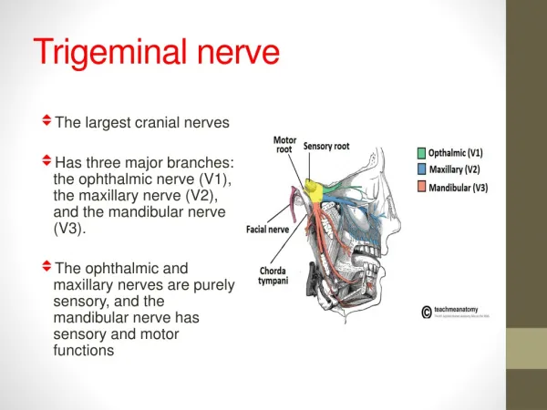
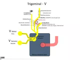
Trigeminal Nerve(V) • Mixed nerve • Largest of the cranialnerves • Threebranches: • Ophthalmic nerve (GSA) ← superior orbital fissure • Maxillary nerve (GSA)← foramen rotundum ← pterygopalatine fossa • Mandibular nerve (GSA, SVE) ← foramen ovale ← infratemporal fossa trigeminal ganglion Pons
Trigeminal Nerve(V) • Emerges from two roots on the ventrolateral surface of thepons • The large sensory root (GSA) ‐ trigeminal ganglion in the trigeminal cave of the duramatter • The small motor root (SVE) originates from the pons pass beneath theganglion • Join the mandibular branchfor • mastication
Trigeminal NerveNuclei • Motor nucleus of trigeminal nerve(SVE) • Location –pons • Connections • Cortex • Reticular formation, red nucleus,tectum • Fibers course
Trigeminal Nerve SensoryNuclei • Main sensory nucleus(GSA) • Touch & pressure • Location,Extention • Relation to • Motornucleus • Spinalnucleus • Spinal nucleus*(GSA) • Pain & tempreture • Location • Extentions • Medulla –C2 • Somatotopicorganisation • Ophthalmic – mostcaudal • Mandibular – mostrostral • *receive GSA from other cranial nerves • Mesencephalic nucleus(GSA) • Proprioception • Location,Extension
Trigeminal Nerve SensoryNuclei • 1st orderneurons • Trigeminal ganglion • 2nd orderneurons • Trigeminal nerve sensory nuclei • Central axons crossmidline • Form trigeminallemniscus • 3rd orderneurons • VPM nucleus ofthalamus • Internal capsule
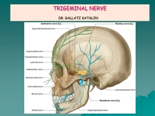
Maxillary Nerve:Branches • Menengialbranches • Zygomatic branch – inferior orbital fissure • Zygomaticotemporal n.‐ • Zygomaticotemporalforamen • Zygomaticofacial n. ‐ Zygomaticofacial foramen • Posterior superior alveolar n.– • posterior superior alveolarforamen • Infraorbital nerve – inferior orbital fissure – infraorbital groove – infraorbitalforamen • Middle superior alveolarn. • Anterior superior alveolarn.
Maxillary Nerve:Branches • Ganglionic branches (pterygopalatinenerves) • Greater &lesser palatine nn. – Greater & lesser palatine canals/foramens • Nasal branches (posterior superior lateral, posterior superior medial, & nasopalatine nn.) – sphenopalatineforamen • Pharyngeal branch – pharyngeal canal
Mandibular Nerve:Branches • Meningeal branch– • foramenspinosum • Nerve to medialpterygoid • Anteriordivision • Massetericn. • Deep temporalnn. • Nerve to lateralpterygoid • Buccalnerve
Mandibular Nerve:Branches • Posteriordivision • Auriculotemporaln. • Relations – TMJ, middle menengeal a. • Lingualn. • Relations – submandibularduct • Inferior alveolar n. – mandibular foramen • Mylohyoid n. (GSE) • Mental n. – mentalforamen
Ophthalmic Nerve:Branches • Superior orbitalfissure • Frontalnerve • Scalp • Branches: • Supraorbital & Supratochlearnn. • Lacrimalnerve • Lateral part of uppereyelid • Carry parasympathetic fibers to lacrimal gland via zygomaticotemporalnerve
Ophthalmic Nerve:Branches • Nasociliary nerve‐Branches: • Comunicating branch to ciliary ganglion‐ sensory fibers from short ciliarynn. • Long ciliary nn.‐ carry sympathetic fibers (dilator pupillaem.) • Posterior ethmoidal n. (ethmoid & sphenoidsinuses) • Anterior ethmoidaln. • External nasal branch (tip ofnose) • Infratrochlear n. (medial part of upper eyelid & part ofnose)
Trigeminal Nerve (V):Lesion • Loss of general sensation (hemianesthesia) from face and oral & nasalcavities • Loss of corneal reflex (V1) (afferentlimb) • Paralysis of the muscles ofmastication • Deviation of the mandible to the weakside • Paralysis of the tensor tympani muscle – partial deafness to low‐pitchedsounds • Trigeminalneuralgia • Test • Sensory – by touching face using cottonball • Motor – by assisting masticatory muscles (masseter & temporalis) onclenching