Download
1 / 25
320 likes | 1.01k Vues
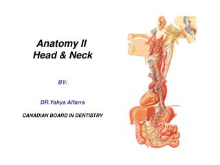
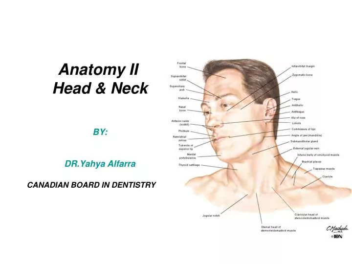
Anatomy II Head & Neck BY: DR.Yahya Alfarra CANADIAN BOARD IN DENTISTRY. Superficial Anatomy of the Neck. The neck lies between The lower margin of the mandible. The suprasternal notch and upper border of the clavicle. Fascia of the neck. Superfiscial fascia :

E N D
Anatomy II Head & Neck BY: DR.Yahya Alfarra CANADIAN BOARD IN DENTISTRY
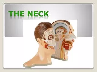
Superficial Anatomy of the Neck The neck lies between • The lower margin of the mandible. • The suprasternal notch and upper border of the clavicle.
Fascia of the neck • Superfiscial fascia : A thin layer immediately beneath the skin and encloses: • The platysma muscle • Cutaneous nerves • Superficial veins : External Jugular Vien. • Superficial lympatics • Deep fascia : • Investing layer • Pretracheal layer • Prevertebral layer • Carotid sheath
Cutaneous Nerves of the Neck Anteriolateral region : • Lesser occipital nerve (C2) • Greater auricular nerve (C2, C3) • Transverse cutaneous nerve(C2, C3) • Supraclavicular nerves(C3, C4) (medial, intermediate, lateral).
Cutaneous Nerves of the Neck Ant. Lateral area: Posterior region: over the trapezius m. and the posterior scalp . • Greater occipital nerve (post ramus of C2) • Posterior rami of C3, C4 and C5.
Platysma • Origin: deep fascia that covers the upper parts of the pectoralis major and deltoid muscles • Insertion: lower margin of the mandible and the angle of the mouth • Innervation: cervical branch of the facial nerve (CN VII) • Action: depresses the mandible and the lower lip and the angle of the mouth.
Cutaneous Nerves of the Neck • Trigeminal N and it’s branches: • Ophthalmic N. • Maxillary N. • Mandibular N.
Superficial veins of the neck The external jugular vein • It begins by the union of of two veins: 1. Posterior auricular vein 2. A branch of the retromandibular (posterior facial) vein.
Anterior jugular vein • Starts below the chin by the union of small veins • Unite in the suprasternal notch forming the jugular arch
Triangles of the neck • Anterior triangle(by hyiod Bone divide into 4 triangles By 2 Ms ( Digastric Ms+omohyoid Ms) • Submental • Digastric • Carotid • Muscular • Posterior triangle • Occipital • Supraclavicular (Subclavian triangle)
Anterior triangle of the Neck • Boundaries: • Midline • Anterior border of SCM • Lower margin of the mandible subdivided into • 4 triangles by 2Ms • Digastric Ms. • Omohyoid Ms. • (topography)
Posterior triangle of the Neck • Posterior triangle • Occipital • Supraclavicular (Subclavian triangle) Borders: *SCM *Trapezius Ms *Middle third of clavicle
Nerves of post. triangle 1- spinal accessory N.?? 2- Cervical plexus 3- Brachial plexus
Sternoclidomastoid Muscle • Origin: - sternal head: manibrium sterni - clavicular head: medial upper third of clavicle • Insertion: The two heads unite and insert into the mastoid tip and superior nuchal line
Nerve supply: Spinal root of accessory nerve • Action: Extention of head & Flextion of neck Note: N. crossing SCM. ??
Omohyoid muscle • Inferior belly: originates from the upper margin of the scapula, passes deep to the SCM and ends in the intermediate tendon. • Intermediate tendon: held by the deep fascia of the clavicle and first rip. • Superior belly: inserts into the lower border of the hyoid bone body. • Nerve supply: Ansa cervicalis (C1, 2, 3) • Action: depresses the hyoid bone
Strap muscles(Infrahyoid muscles) 1-Thyrohyoid -- deepest 2-Sternothyroid _ deepest 3-Sternohyoid 4-Omohyoid • Together with the suprahyoid muscles stabilize the hyoid bone to provide movements of the tongue and the larynx during swallowing.