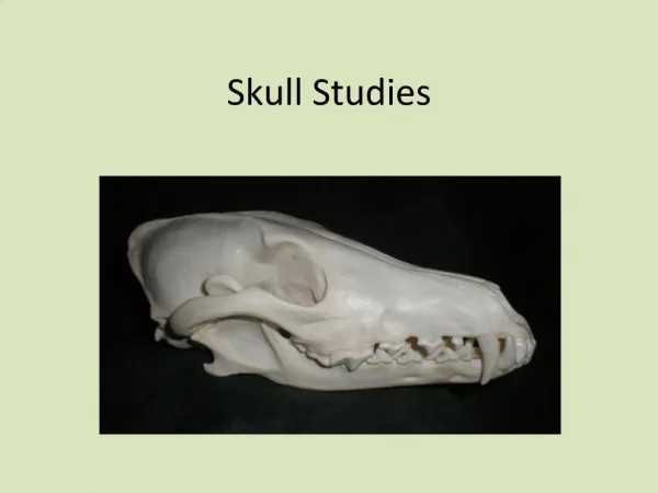
Skull
Skull. Objectives. Identify the different bones forming the skull with important foramina . Differentiate between the bony features of the skull of an adult and infant. The Skull. It consists of flat and irregular bones Divides into: Cranial bones which encase the brain

Skull
E N D
Presentation Transcript
Objectives • Identify the different bones forming the skull with important foramina. • Differentiate between the bony features of the skull of an adult and infant.
The Skull • It consists of flat and irregular bones • Divides into: Cranial bones which encase the brain Facial bones which form the bones of the face • The following must be identified: Nasion Inion Bregma Lambda External occipital protuberance Parietal eminence Zygomatic arch
The Skull • Facial bones include: (Viscerocranium) Maxilla Zygomatic Nazal Palatine Lacrimal Inferior concha Vomer Mandible • Cranial bones include: (Neurocranium) Frontal Ethmoid Sphenoidal Occipital Parietal Temporal
The Skull • Frontal • Maxilla • Zygomatic • Nazal • Mandible • Lacrimal
The Skull • Parietal • Temporal Squamous Mastoid Styloid Tympanic Petrous • Sphenoid Greater wing Lesser wing Pterygoid process Body • Occipital
The Skull • Petrous part of temporal • Lesser wing of sphenoid • Body of sphenoid • Pterygoid process • Ethmoid bone Crista galli Cribriform plate Perpendicular plate • Vomer • Palatine bone
Cranial Fossa • It is the cavity of the skull • Divides into: Anterior cranial fossa Middle cranial fossa Posterior cranial fossa
Anterior Cranial Fossa • Formed of: Orbital plate of frontal bone Cribriform plate of ethmoid Crista galli Lesser wing of sphenoid • Occupies: Frontal lobe of the cerebrum Optic nerves Olfactory bulb and tract
Middle Cranial Fossa • Formed of: Greater wing of sphenoid Body of sphenoid Squamous part of temporal bone Anterior part of petrous bone • It occupies: Temporal lobe of the cerebrum
Posterior Cranial Fossa • Formed of: Sphenoid part of clivus Posterior part of petrous bone Occipital bone • It shows the following land marks: Groove for transverse sinus Groove for segmoid sinus Internal occipital protuberance • It occupies: Cerebellum Brain stem
Base of The Skull • Most of the bones are seen in other views • Fracture base of the skull why?
Foramina of the Skull • Foramina are found within the skull bones • They vary in size and shape • They transmit nerves, blood vessels and dura matter
Foramina of the Skull • Supraorbital • Infraorbital • Mental
Sutures of the Skull • Sutures are fibrous joints articulating the skull bones • They include: Coronal Sagittal Lambdoid Pterion
Skull Fontanel • Fontanels are spaces between some of the skull bones of infants • They permit easy growth of the brain • They permit overlapping of skull bones during labor • They are anterior, posterior, anterolateral, and postero- lateral • They close later in life at 6-18 months
The Mandible • It is a flat bone • Identify the following: Head(condylar process) Mandibular notch Coronoid process Ramus Angle Body Mylohyoid line Submandibular fossa Digastric fossa Mandibular foramen Mental foramen