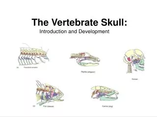
The Skull
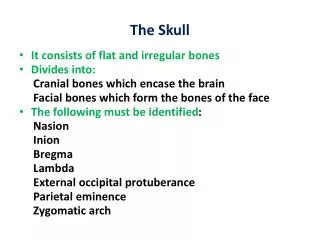
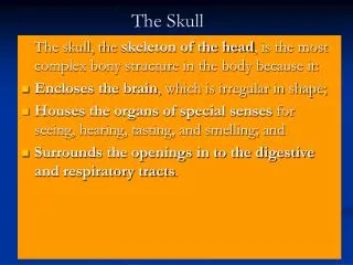
The Skull. The skull, the skeleton of the head , is the most complex bony structure in the body because it: Encloses the brain , which is irregular in shape; Houses the organs of special senses for seeing, hearing, tasting, and smelling; and

The Skull
E N D
Presentation Transcript
The Skull The skull, the skeleton of the head, is the most complex bony structure in the body because it: • Encloses the brain, which is irregular in shape; • Houses the organs of special senses for seeing, hearing, tasting, and smelling; and • Surrounds the openings in to the digestive and respiratory tracts.
The Skull • In the anatomical position, the skull is oriented so that the inferior margin of the orbit (eye socket) and the superior margin of the external acoustic meatus (auditory canal) are horizontal. This is called the orbitomenial plane (Frankfort plane).
the cranium is often used when referring to the part of the skull containing the brain. Skull is the collection of all bones including the cranium along with mandible and all facial bones
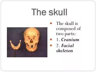
The skull is comprised of many bones that are closely fitted together. A series of flat bones are united by interlocking sutures to form the calvaria, and a group of irregular bones form the face and the cranial base. The skull as a whole is of greater importance to most health professionals than are its constituent bones, but it is important to understand how the skull is constructed. Bones of the Skull
Bones of the Skull • Except for the mandible rigid sutures join the bones of the adult skull. The cranium is essentially a single complex bone. • Although the adult skull is rigid, the bones forming it in infants and children grow as individual bones undergo remodelling. Furthermore, relationships among the various bones are constantly changing during these developmental periods.
The Frontal Bone Bones of the Calvaria
The frontal bone forms the thin roof of the orbits (eye sockets). • Just superior to and parallel with each supraorbital margin is a bony ridge, the superciliary arch, which overlies the frontal sinus. This arch is more pronounced in males.
The frontal bone articulates with the two parietal bones • And the nasal bones medially • The frontal bone also articulates with the zygomatic, laterally • In about 8% of adult skulls, a remnant of the inferior part of the metopic (interfrontal) suture is visible. It may be mistaken in radiographs for a fracture line by inexperienced observers.
The superciliary arches are relatively sharp ridges of bone and a blow to them may lacerate the skin and cause bleeding.
The Parietal Bones • The two parietal bones (L. paries, wall) form large parts of the walls of the calvaria. • Forms sides and roof of the cranium
The parietal bones articulate with each other in the median plane at the sagittal suture. The medial plane of the body passes through the sagittal suture. • The inverted V-shaped suture between the parietal bones and the occipital bones is called the lambdoid suture because of its resemblance to the letter lambda in the Greek alphabet. • The point where the parietal and occipital bones join is a useful reference point called the lambda. It can be felt as a depression in some people.
The Temporal Bones • The sides and base of the skull are formed partly by these bones.
The temporal bone articulates at sutures with the parietal, occipital, sphenoid, and zygomatic bones. • The zygomatic process of the temporal bone unites with the temporal process of the zygomatic bone to form the zygomatic arch. The zygomatic arches form the widest part of the face. • The head of the mandible articulates with the mandibularfossa on the inferior surface of the zygomatic process of the temporal bone. • Because the zygomatic arches are the widest parts of the face and are such prominent facial features, they are commonly fractured and depressed. A fracture of the temporal process of the zygomatic bone would likely involve the lateral wall of the orbit and could injure the eye.
Fetal Skull An infant's skull is composed of 6 separate bones (the frontal bone, the occipital bone, 2 parietal bones, and 2 temporal bones), called cranial bones. These bones are held together by strong, fibrous, elastic tissues called cranial sutures. The spaces within the fibrous tissues between the bones (sometimes referred to as "soft spots") are called Fontanelles (the anterior fontanelle and the posterior fontanelle). The cranial bones remain separate bones for approximately 12 to 18 months. Then the separate cranial bones grow together (fuse) and remain fused throughout adulthood.
The large fibrous area where several sutures meet are called fonticuli or fontanelles. • The softness of these bones and looseness of their connections at these sutures enable the calvaria to undergo changes of shape during birth called molding. Within a day or so after birth, the shape of the infant’s calvaria returns to normal. • The loose construction of the new-born calvaria also allows the skull to enlarge and undergo remodelling during infancy and childhood. Relationships between the various bones are constantly changing during the active growth period. • The increase in the size of the cranium is greatest during the first 2 years, the period of most rapid postnatal growth of the brain. • The cranium normally increases in capacity until about 15 or 16 years of age; thereafter the cranium usually increases only slightly in size as its bones thicken for 3 to 4 years.
During infancy and childhood, the flexibility of the fibers allows the rapid growth of the brain without constriction while protecting the brain from minor impacts to the head (such as when the infant is learning to hold his head up, roll over, and sit up). Without the flexibility of the sutures and fontanelles, the child's brain would be constricted within the cranial bones and could not grow adequately. The child would suffer brain damage.
Feeling the cranial sutures and fontanelles is one way that physicians and nurses determine the child's growth and development. They are able to assess the pressure within the brain by feeling the tension of the fontanelles. The fontanelles should feel flat and firm. Bulging fontanelles indicate increased pressure within the brain. In this case investigation with imaging techniques such as CT scan or MRI scan is warranted and surgery may be necessary to relieve the increased pressure. Sunken, depressed fontanelles indicate dehydration
Fetal skull • Fontanelles • Unossified membranous gaps between the skull bones in fetus. • Anterior. • Posterior
Fontanellaes • Fontanelles are the "soft spots" on an infant’s head where the bony plates that make up the skull have not yet come together. It is normal for infants to have these "soft spots", which can be seen and felt on the top and back of the head. Fontanelles that are abnormally large may indicate a medical condition.
Anterior Fontanelle Bregma the meeting point of sagittal and coronal sutures between frontal and parietal bones,fuses by 1.5 yrs of age
Posterior Fontanelle Lambda , the meeting point between sagittal and lambdoid sutures, corresponding to postierfontanelle, fuses at 2-3 months of age
Occipital bone • The occipital bone forms the back part of the skull and the base of the cranium • It joins with the parietal and temporal bones. • In the center, underside (inferior) portion of the cranium, there is a large opening called the foramen magnum • It transmits the medulla oblongata and its membranes
Zygomaticbone • The zygomatic bone is small and quadrangular, and is situated at the upper and lateral part of the face: it forms the prominence of the cheek, part of the lateral wall and floor of the orbit • The temporal process, long, narrow, and serrated, articulates with the zygomatic process of the temporal.
The frontosphenoidal process is thick and serrated, and articulates with the zygomatic process of the frontal bone. • The orbital process is a thick, strong plate, projecting backward and medialward from the orbital margin.