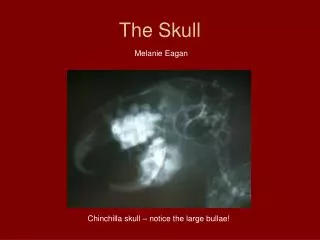
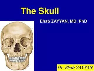
The Skull
The Skull. 王 配 军. http://anatomy.doclink.cn http://www.yymc.edu.cn/yuanxi/jcb/jpk/jpk-index.htm E-mail:peijunwang568@yahoo.com.cn. 目的要求. 1. 掌握颅的组成,脑颅、面颅的划分。 2. 握颅底内面观的主要结构。 3. 掌握眶、骨性鼻腔的形态特征。骨性鼻旁 窦的名称、位置及开口。 4. 掌握颅骨的常用骨性标志:眉弓、颧弓、 下颌角、舌骨、乳突、枕外隆突。 5. 了解新生儿颅骨的特征及生后变化。. The skull.


The Skull
E N D
Presentation Transcript
The Skull 王 配 军 http://anatomy.doclink.cn http://www.yymc.edu.cn/yuanxi/jcb/jpk/jpk-index.htm E-mail:peijunwang568@yahoo.com.cn 郧阳医学院解剖教研室
目的要求 1.掌握颅的组成,脑颅、面颅的划分。 2.握颅底内面观的主要结构。 3.掌握眶、骨性鼻腔的形态特征。骨性鼻旁 窦的名称、位置及开口。 4.掌握颅骨的常用骨性标志:眉弓、颧弓、 下颌角、舌骨、乳突、枕外隆突。 5.了解新生儿颅骨的特征及生后变化。 郧阳医学院解剖教研室
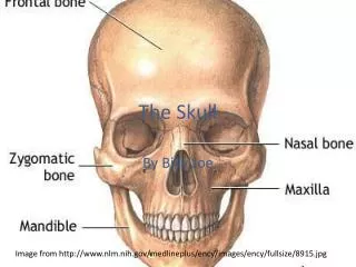
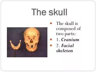
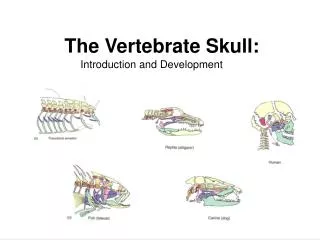
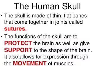
The skull The skull contains 23 bones .The skull is divided into two parts :cerebral cranium and facial Cranium. 郧阳医学院解剖教研室
(一)The cerebral cranium The cerebral cranium consists of eight cranial bones . one frontal one ethmoid one sphenoid one occipital two parietal two temporal 郧阳医学院解剖教研室
(二)The facial cranium The facial cranium are fifteen in number. maxilla palatine paired zygomatic bones nasal lacrimal bones inferior nasal conchae vomer Unpaired mandible bones hyoid 郧阳医学院解剖教研室
1. The mandible angle of mandible 2. The hyoid 郧阳医学院解剖教研室
(三)The skull as a whole 1.The superior aspect of the skull coronal suture sagittal suture lambdoid suture 2.The posterior aspect of the skull external occipital protuberance superior nuchal line 郧阳医学院解剖教研室
3.The internal surface of the base of skull It is divided into three fossa. These are the anterior,the middle and the posterior cranial fossa. 郧阳医学院解剖教研室
(1) Theanterior cranial fossa frontal crest foramen cecum crista galli cribriform plat cribriform foramina 郧阳医学院解剖教研室
(2) Themiddle cranial fossa hypophysial fossa optic canal anterior clinoid process dorsum sellae posterior clinoid process superior orbital fissure foramen lacerum foramen rotundum foramen ovale foramen spinosum sulcus for middle meningeal artery 郧阳医学院解剖教研室
(3) Theposterior cranial fossa foramen magnum hypoglossal canal internal occipital protuberance sulcus for superior sagittal sinus sulcus for transverse sinus sulcus for sigmoid sinus jugular foramen internal acoustic pore 郧阳医学院解剖教研室
4.The external surface of the base of skull alveolar arch incisive foramina greater palatine posterior nasal apertures occipital condyle hypoglossal canal jugular foramen styloid process stylomastiod foramen 郧阳医学院解剖教研室
5.The lateral view of skull external acoustic pore mastoidprocess zygomatic arch temporal fossa infratemporal fossa pterion infratemporal fossa pterygopalatine fossa 郧阳医学院解剖教研室
翼点 硬膜外出血 郧阳医学院解剖教研室
6.The front view of skull (1) the forehead superciliary arch (2) the orbits ① orbital opening supraorbital foramen (notch) ② orbital apex:optic canal ③ superior wall:lacrimal fossa ④medial wall: lacrimal sac fossa, nasolacrimal canal ⑤ inferior wall: inferior orbital fissure, inferior orbital foramen ⑥ lateral wall: superior orbital fissure 郧阳医学院解剖教研室
(3) the bony nasal cavity nasal septem superior middle conchae inferior superior middle meatus inferior sphenoethmoidal recess 郧阳医学院解剖教研室
(4) the paranasal sinuses ① the frontal sinuses ② the ethmoidal sinuses ③ the sphenoidal sinuses ④ the maxillary sinuses 郧阳医学院解剖教研室
(5) the oral cavity (四)The skull at birth 郧阳医学院解剖教研室
小 结 郧阳医学院解剖教研室
Thank you ! 郧阳医学院解剖教研室