PULMONARY HYPERTENSION
PULMONARY HYPERTENSION. Prof. S. Shanmuga Sundaram K.S. Hospital, Chennai. PULMONARY HYPERTENSION AT SYSTEMIC LEVEL, DUE TO HIGH PULMONARY VASCULAR RESISTANCE ( > 800 dynes sec cm -5 ) WITH REVERSED OR BIDIRECTIONAL SHUNT…..

PULMONARY HYPERTENSION
E N D
Presentation Transcript
PULMONARY HYPERTENSION Prof. S. Shanmuga Sundaram K.S. Hospital, Chennai
PULMONARY HYPERTENSION AT SYSTEMIC LEVEL, DUE TO HIGH PULMONARY VASCULAR RESISTANCE ( > 800 dynes sec cm-5 ) WITH REVERSED OR BIDIRECTIONAL SHUNT….. 8% (1950) → → → → 4%
DEATH: Sudden death 30% , Heart failure 23% , Hemoptysis 11% Death during noncardiac surgery & pregnancy
DISSECTION OF PULMONARY ARTERY PROXIMAL PA THROMBOSIS
PULMONARY ARTERIAL HYPERTENSION IN SHUNT LESIONSmPAP > 25 mmHg at rest / > 30 mm Hg post exercisePAWP < 15 mm Hg ; PVR > 3 Wood Units • TRANSMISSION OF SYSTEMIC ARTERIAL PRESSURE • VASOCONSTRICTION • VASCULAR OBLITERATION – MEDIAL HYPERTROPHY INTIMAL PROLIF + FIBROSIS ARTERIAL THROMBI HYPERKINETIC OBLITERATIVE PVR < 5 W.U > 5 W.U PA PP/PA SP > 60% < 40%
ASD VSD PDA CA, APVC TGA VSD, DORV APWINDOW SINGLE VENTRICLE TRUNCUS > 2 cm > 1 cm > 1 cm
PULMONARY CIRCULATION - STRUCTURAL REMODELING Elastic > Fully muscular > Partially muscular > Non muscular • At birth the smallest muscular arteries dilate with medial thinning • By 4 months, this process involves larger arteries & get completed • Alveoli and Arteries grow both in number & size Al : Art = 20:1 > 8:1 • With shunt lesions resulting in increased flow ± pressure, proximal arteries dilate, distal arteries reduce in number and size bcause of extension of muscle in media of partially or non muscular arteries
PAH IN L > R SHUNTS • NONRESTRICTIVE VSD = 15 % < 2 yrs of life MODERATE DEFECTS = 3% ; LARGE DEFECTS (1.5cm) = 50% • LARGE PDA = similar incidence • LARGE ASD = 6-10% > 3rd decade Frequent in SVC, partial AV Canal defects & in Lutembacher’s • TGA = 8% (intact IVS) 40% ( VSD/PDA ) < 1 yr 75% at 2 yrs • COMMON AV CANAL all develop PAH • TRUNCUS ARTERIOSUS by 1-2 yrs • SYSTEMIC - PA SHUNTS: BT Shunt (<10%) Waterston / Pott’s ~ 30%
MECHANISMS OF PAH IN L>R SHUNTS LESION ↑Qp ↑PAP ↑PVP ↓ pH ↑ Ht ASD + - - - - VSD + + + - - PDA + + + - - AV CANAL + + ++ - - TGA, TA + + + + +
ENDOTHELIAL DYSFUNCTION ↑ ET, TXA2 , SEROTONIN ↓ NO ,PGI2,VIP PLATELET ADHESION + THROMBUS
GENETIC SUSCEPTIBIILITY BMPR2 MUTATION = 6 % 26%(IPAH) 50% (FPAH)
MORPHOMETRIC GRADING Rabinovitch M Grade A : Extension of muscle into small peripheral arteries Wall thickness increased but < 1.5 times the normal ↑ PBF ↑ PA PP + NORMAL MEAN PAP Grade B : Mild : medial thickness 1.5 – 2.0 times the normal Severe : medial thickness > 2 times the normal PAH - MEAN PAP > 50 % OF SYSTEMIC LEVEL Grade C : Size and number of arteries reduced PAH - PVR > 3.5 - 6.0 u.m2
CLINICAL RECOGNITION • Apparent improvement of neonatal HF • Reduction of frequency of respiratory infections • Precordium becomes less tumultous • Flow murmur decreases > disappears • Shunt murmur decreases in intensity & duration • S2 split decreases and P2 increases in intensity
EISENMENGER’S SYNDROME SYMPTOMS: 1) Low C.O + Hypoxia > DOE, Dizziness, Syncope, Fatigue 2) Hemoptysis: Rupture of plexiform, dilatation lesions, pulmonary arterioles, Broncho Pulmonary connexions, Pulmonary Embolism / in situ thrombosis 3) Hyperviscosity: Headache, dizziness, Visual sx 4) Right Heart failure : Edema, RHC pain 5)CVA :Hyperviscosity, Parad. emboli, Cerebral abscess 6) Sudden cardiac death: Arrhythmia
EISENMENGER’S SYNDROME SIGNS : 1) Cyanosis and Clubbing 2) JVP inconspicuous 3) Pulmonary Ejection Sound 4) 2-3/6 Ejection Systolic Murmur 5) Loud P2 6) Murmurs of TR and PR
EISENMENGER’S SYNDROME FEATURE ASD VSD PDA Neonatal HF - + + Age 30-40 2-12 2-12 Syncope ± ± - Cyanosis Uniform Uniform Differential Cardiomegaly,PSH + - - Wide pulse pressure - - ± Prominent ‘a’ JVP + - - S2 split Fixed Single Normal Long PR murmur - - +
PDA DOPPLER PATTERNS PAH GROWING PULSATILE CLOSING CLOSED
SHUNT LESIONS - OPERABILITY • Qp : Qs = > 2:1 No or mild PAH • Qp : Qs = < 1.5:1 Severe PAH - INOPERABLE Qp = O2 Consumption / PV – PA O2 content Qs = O2 consumption / SA - MV O2 content O2 content = O2 saturation x O2 carrying capacity x Hb Qp : Qs = SA – MV O2 sat / PV – PA O2 sat
CHD PAH – REVERSIBILITY TESTING HIGH SURGICAL RISK ( 20% ) RIGHT VENTRICULAR FAILURE ( IPAH like ! ) PROGRESSION OF PAH AGENTSCRITERIA 100% OXYGEN (10 mts) ↓Rp /Rs > 20% NITRIC OXIDE (10-80ppm) Rp:Rs < 0.33 02 + N.O (Se 97% Sp 90%) Rp < 8 u.m2 ADENOSINE (50-500µg/kg/mt) EPOPROSTENOL (2-10 ng/kg/mt) ILOPROST (2.5-5.0 µg )
ASSESSING OPERABILITY BASED ON PVRMISTAKES & MISCONCEPTIONSExpecting PAP to decline ( ↓ PVR > ↑ FLOW )Assuming O2 consumptionIgnoring dissolved O2 in calculating PVRO2 sat x 1.36 x Hb = 60 x 1.36 x 12 = 98 ml/L ( 0.03 x 55 = 1.7ml ) 98 x 1.36 x 12 = 160 ml/L ( 0.03 x 95 = 2.9ml ) PVR = 60 – 8 = 52 / 3.2 = 16 units ( 16.5 units ) After 100% oxygen : 72 x 1.36 x 12 = 118 ml/L ( 0.03 x 100 = 3 ml ) 98 x 1.36 x 12 = 160 ml/L ( 0.03 x 500 = 15 ml) PVR = 55 – 8 = 47 / 4.8 = 9.8 units ( 12.7 units ) 22 to 44% 40 to 60% 60 to 100%
PVR INDEXED TO BODY SURFACE AREA A child of BSA of 0.5 m2 has a PBF of 2 l/mt PA mean pressure = 20 mmHg ; mean LAP = 8 mmHg PVR absolute value = 20-8/2 = 6 units If corrected for BSA = 6/0.5 = 12 units PBF corrected to BSA = 2/0.5 = 4 l/mt/m2 PVR indexed to BSA = 20-8/4 = 3 u.m2
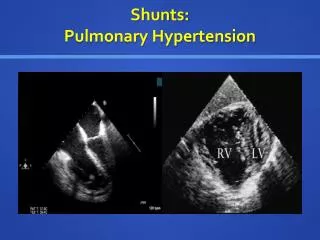
ROLE OF ECHOCARDIOGRAPHY • Qp/Qs by doppler, PAcT not reliable • PA peak velocity > 1.0 m/s predictive • PVR = TR Velocity/ TVI RVOT x 10 + 0.16 • Vp > 18 cm/s = PVR < 6 units 8.8 W.U 4 WU 16.4 W.U 12.4 cm/s 23.1 cm/s
PREDICTION OF PVODWilson NJ CCVD 1993;28:22 PREDICTING HEATH EDWARDS Grade III - IV Sensitivity Specificity PVR > 6 units 100% 94% Monopedial count<3 83% 100% Abnormal blush 83% 69% Combination of all 100% 100%
LUNG BIOPSYMORPHOMETRIC GRADING Rabinovitch M Grade A : Extension of muscle into small peripheral arteries Wall thickness increased but < 1.5 times the normal ↑ PBF ↑ PA PP + NORMAL MEAN PAP Grade B : Mild : medial thickness 1.5 – 2.0 times the normal Severe : medial thickness > 2 times the normal PAH - MEAN PAP > 50 % OF SYSTEMIC LEVEL Grade C : Size and number of arteries reduced PAH - PVR > 3.5 - 6.0 u.m2
CARDIAC MR • DEFECT SIZE & LOCATION • PA SIZE ↑ WITH PAH • RV FUNCTION • Qp/Qs RATIO Phase contrast velocity mapping • MR OXIMETRY ( T2 relaxation time) • DEGREE OF PAH
BALLOON OCCLUSION IN HYPERTENSIVE DUCTUSRoy A IHJ 2005;57:332 Fall in m/d PAP > 20 mmHg
TRIAL OCCLUSION OF PDA Yan C Heart 2007;93:514 Trial occlusion for 30 mts with ADO Reduction of mPAP 78 ± 19.3 to 41 ± 13.8 mm Hg FU for 3 to 6 months – clinical improvement
PAH IN ATRIAL SEPTAL DEFECT • 6% ( Mayo clinic); 9% - half were below 20 yrs(CMC) • PAH (mPAP>30 mmHg) 26% SVC (9% FO) ↑PVR 16% SVC (4% FO ) ; at younger age • 85 % were women ( overall F:M = 2:1 ) • PVR > 15 units do poorly – death / progression of PAH • PVR < 10 units do well with surgery • PVR 10 – 15 units – if SPO2 is < 90% surgery not useful
DEVICE CLOSURE IN ASD + PAHBalint OH Heart 2008;94:1189 PAH Moderate Severe PASP 50-59 >60 At 3 m PASP ↓ 57± 11 to 51±17 At 3 yrs PASP ↓ to 44 ±16 Only in 43.6% PAP normalised 15.4% had persistent severe PAH
EISENMENGER’S SYNDROMEMANAGEMENT ISSUES • Avoid dehydration, living at high altitude • Air travel safe (supplemental O2) • Avoid pregnancy ( No OCP – tubal ligation/vasectomy) • Treat Iron deficiency ( MCV < 82 ) ; hyperuricemia • Vensection for hyperviscosity syndrome • Antiplatelet / Anticoagulants ? • Disease targeting therapies : Prostacyclin & analogues, sildenafil, bosentan • Surgery: Correction after PA banding, prolonged vasodilator therapy, Heart Lung Transplant
PROGNOSIS EISENMENGER SYNDROME ~ IPH ACTUARIAL SURVIVAL E.S IPAH 1 yr 97 % 77 % 2 yr 89 % 69 % 3 yr 77 % 35 %
MORPHOMETRIC GRADING Rabinovitch M Grade A : Extension of muscle into small peripheral arteries Wall thickness increased but < 1.5 times the normal ↑ PBF ↑ PA PP + NORMAL MEAN PAP Grade B : Mild : medial thickness 1.5 – 2.0 times the normal Severe : medial thickness > 2 times the normal PAH - MEAN PAP > 50 % OF SYSTEMIC LEVEL Grade C : Size and number of arteries reduced PAH - PVR > 3.5 - 6.0 u.m2