Pulmonary Hypertension
Pulmonary Hypertension. Allison K. Cabalka, MD Associate Professor of Pediatrics Consultant, Pediatric Cardiology Mayo Clinic. CHD and PHTN: Issues. What is Pulmonary HTN? How does one define elevated pulmonary artery pressure Classification scheme How do we assess PHTN in those with CHD?

Pulmonary Hypertension
E N D
Presentation Transcript
Pulmonary Hypertension Allison K. Cabalka, MD Associate Professor of Pediatrics Consultant, Pediatric Cardiology Mayo Clinic
CHD and PHTN: Issues • What is Pulmonary HTN? • How does one define elevated pulmonary artery pressure • Classification scheme • How do we assess PHTN in those with CHD? • Non-invasive: clinical, echo • Invasive: cath evaluation • Treatment of PHTN?
What is Pulmonary HTN? • Simple Description Elevated PA pressure ( pulm vein pressure) • Definition (mm Hg) PA pressure >30/15 PA (mean) >25 Mild 26-35 Moderate 36-50 Severe >50
Why is PTHN Important? • It is a disease of the entire pulmonary circulation • It is a critical determinant of outcome • Morbidity, mortality in pediatric lung, cardiac, pulmonary, hematologic, and other diseases
Evian-Venice Classification • PHTN in the setting of CHD with systemic to PA shunts • Classified as Pulmonary Arterial Hypertension • Considering the following: • Type of CHD • Prognosis/evolution of pulmonary vascular disease • Circulatory physiology World Symposiums on PHTN; Evian 1998 and Venice 2003
Characterizing PHTN in CHD • Presence of systemic-to-pulmonary shunt • Location of shunt • Direction of shunt • Size of defect • State of repair • Associated cardiac anomalies Evian/Venice Classification
Systemic-to-Pulmonary Shunt • Yes/No • Previously present by history? • If not, consider another classification system (such as WHO) • For purposes of this discussion, we will assume all pts have a shunt lesion (CHD) Evian/Venice Classification
Location of Shunt? • Pre-tricuspid level • Inter-atrial communication (ASD) • Anomalous pulmonary venous drainage/connection • Post-tricuspid level • Ventricular septal defect (VSD) • Patent ductus arteriosus/AP window • Functionally univentricular hearts Evian/Venice Classification
Direction of Shunt? • Systemic to pulmonary • Left-to-right • Pulmonary to systemic • Right-to-left • Bidirectional Evian/Venice Classification
Size of Defect? • Anatomic and functional • Consider size at presentation and current size • Quantification of shunt • Ratio of pulmonary and systemic flows • Restriction? • Is there any pressure gradient through the post-tricuspid defect? Evian/Venice Classification
State of Repair? • Unoperated • Palliated • Age at repair • Type of surgery • Repaired • Age at repair • Type of surgery Modified from Venice 2003
IF Postoperative Patient? • Correction of shunt and age at correction • Pulmonary arterial banding • Age at PAB, duration of banding • Presence and type of surgical shunts • Blalock-Taussig, Pott’s, Waterston • Residual shunting • Quantification and direction Evian/Venice Classification
Associated Cardiac Anomalies Especially those that affect pulmonary hemodynamics: • Pulmonary valve stenosis • Defects affecting pulmonary venous “outflow” • Cor triatriatum, mitral stenosis, LV dysfunction • Ventricular function • Systolic, diastolic • Overall cardiac output Evian/Venice Classification
Considering CHD and PHTN Basic issues at presentation: • Is the patient still repairable? • Is the shunt lesion recognized in a timely fashion? • Is the pulmonary hypertension reversible? • There may be a point of “no return” but the time course varies widely from patient to patient…
Pulmonary Arterial HTN In patients with CHD: • Pulmonary artery is exposed to systemic pressure when VSD or PDA is unrestrictive • Pulmonary HTN is present from birth • Pulmonary vascular resistance determines outcome
What is the Point of No Return? Eisenmenger syndrome: • Pulmonary vascular resistance is elevated • Shunt reverses and becomes right-to-left
PVD: Who is at Risk? • Shunt + Pulmonary • Hypertension Pulmonary Vascular Disease Particularly if there is Cyanosis
Two Ventricles… • Large VSD, Large PDA • Combined intracardiac shunts • i.e. d-TGA with VSD; DORV • Cyanosis and mixing is a BAD combination • What about large ASD? • This may be a separate issue • i.e. Primary pulmonary HTN • Other factors: • Pulmonary venous obstruction, arch obstruction (Shone’s syndrome), AV valve regurgitation
One Ventricle… • Basically anyone with an unprotected PA bed (no PS) • Tricuspid atresia with unrestrictive VSD, no PS • DILV without pulmonary stenosis • DORV with small LV, no PS • Ductal-dependent, complex CHD with persistently large PDA
Assessment of PHTN In the Setting of CHD
Assessment in CHD • Early recognition and referral of CHD is critical • This is extremely important! • Natural history may vary; timing or presentation may also vary • Clinical evaluation • Symptoms, growth pattern, other illnesses and hospitalizations? • Physical examination • Laboratory evaluation
Clinical Assessment in CHD Clinical exam: • Does the patient have evidence of large L-to-R shunt? • S2, P2 component • Continuous PDA murmur • Systolic murmur with diastolic flow rumble through mitral valve • Or not? • Loud, single S2 (P2), minimal murmur • Very short systolic murmur • No flow rumble or continuous murmur Is the patient PINK or cyanotic?
CXR in Shunt Physiology This should be reassuring…
CXR in Shunt Physiology This is more reassuring…
CXR in Shunt Physiology This is NOT reassuring…
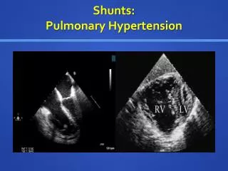
Echo Assessment Careful evaluation of anatomy and physiology: • Confirmation of location, size and associated defects • Doppler profile (velocity) • Shunt direction, left-to-right; bidirectional; right-to-left • Left-sided structural enlargement • Associated valve pathology • Ventricular function
Echo Assessment • Need to be very careful in the patient with a large post-tricuspid defect (i.e. VSD) to NOT miss other defects • Always evaluate the aortic arch and ductus very carefully • Sometimes the PDA with bidirectional shunt is very easy to “miss” • Be careful in the patient with bidirectional or right-to-left shunt • Suspicions of elevated pulmonary resistance? • The patient needs hemodynamic cath study
Shunts and Resistance • Resistance determination is the MAIN reason to cath a child/adult with CHD • Meticulous collection of data! • Resistance is related to pressure divided by flow: Pressure drop across PA bed Rpa = Flow
Looking at Resistance Another Way • Relationship of pressure to flow and resistance PAP = Flow (Qp) • Resistance • IMPT point: Increased pressure can have either or both as possible sources
The “Basic” Cath Study • Hemodynamics needed: • RA and LA (PCWP), MPA mean, and systemic artery mean pressures • Blood samples needed: • Mixed venous (SVC), pulmonary artery, pulmonary vein, aorta • Typically done first in room air • Calculate the Qp, Qs and Resistance • Perform 100% O2 study
Cath: Basic Formulas • Shunts from O2 saturation data • Qp = Pulmonary blood flow (index) • Qp = VO2 / (PV sat-PA sat) (O2 capacity) • Qs = Systemic blood flow (index) • Qs = VO2 / (SA sat- MV sat) (O2 capacity) • VO2 = Oxygen consumption (ml/min/m2) • O2 Capacity = 13.6 x Hgb (gm/dL)
Dissolved Oxygen ? • O2 content = O2 capacity x O2 saturation + PaO2 x .03ml/mmHg • Hgb 11.5 gm/dl • Room air; PV pO2 = 106 • Oxygen content =(11.5x13.6x0.99)+ (106x0.03) = 155 + 3.3 = 158 • 100% Oxygen; PV pO2 = 566, sat 100% • Oxygen content = (11.5x13.6x1.0) + (566x0.03) = 156 + 17 = 173
Dissolved Oxygen? • Remember that Qp = VO2 / (PV sat-PA sat) x (O2 capacity) • If we ignore dissolved O2 then we have a smaller number in denominator, and a “higher” Qp • This will falsely reduce the Rpa in calculations… * Resistance = PAP/ Flow (Qp)
Rpa: Pulmonary Resistance • Normal ≤ 3 wood units•m2 • Borderline between 3-6 wood units•m2 • Over 6 wood units•m2 • Questionable • PHTN may persist or progress • May treat for reversibility over time • Eisenmenger physiology pts usually live longer than those with repair!
Altitude Physiology • Potential contribution of altitude • Delayed transition of decline in pulmonary vascular resistance • Delayed remodeling of vascular bed • Increased muscularization of arteries • Persistence of the PDA • High altitude • Living above 4000 meters • These changes are even more accentuated
Treatment of Elevated Rpa Once you’ve demonstrated that Rpa is elevated… • Treatment with pulmonary vasodilator therapy • Sildenafil • Bosentan • Other more complex regimens are not usually practical
Sildenafil • Cyclic GMP Phosphodiesterase type 5 inhibitor • Prolongs vasodilatory effect of nitric oxide
Sildenafil • Typical adult dose = 20 mg TID • Pediatric dosing • Initial dose: 0.5 mg/kg/dose every 8 hours • Increased if needed and if tolerated • Typical maximum: 2 mg/kg/dose every 6-8 hours
Bosentan • Endothelin receptor antagonist • Endothelin-1 receptors are potent vasoconstrictors; found in higher concentration in lungs of PTHN pts • BREATHE – 5 • Reduced Rpa • Improved 6 min walk distance • With Rx effects may be maintained for 2 yrs (may improve PHTN mortality)
Bosentan • Dosage • 10-20 kg: Initial: 31.25 mg daily for 4 weeks; increase to maintenance dose of 31.25 mg twice daily • >20-40 kg: Initial: 31.25 mg twice daily for 4 weeks; increase to maintenance dose of 62.5 mg twice daily • >40 kg: Initial: 62.5 mg twice daily for 4 weeks; increase to maintenance dose of 125 mg twice daily • Must monitor liver enzymes
Take Home Points • Patient with shunt lesion and obvious increased PBF on clinical/echo evaluation should be repairable • Elevated Rpa on clinical evaluation warrants further study • Cardiac cath and O2 study • Evaluate shunt and reactivity of the pulmonary vascular bed
Take Home Points • Early recognition is critical • If evidence of increased pulmonary resistance consider treatment with pulmonary vasodilator therapy • In patients with obvious Eisenmenger physiology it is better to leave them alone • They will survive longer!!!