Loose Connective Tissue
This overview explores the composition and characteristics of loose connective tissue (LCT), emphasizing areolar tissue and its components. Highlights include collagen, elastin, and proteoglycan structures, with detailed descriptions of major staining techniques such as Masson’s Trichrome, Orecin, and Silver stains. Additionally, the role of fibroblasts, adipocytes, and defense cells like neutrophils and macrophages is examined, showcasing their functions within LCT. Visual aids include transmission electron microscopy (TEM) images, illustrating collagen fibers and other cell types in detail.

Loose Connective Tissue
E N D
Presentation Transcript
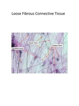
Loose Connective Tissue (LCT)
Areolar Tissue Pink = collagen Purple = elastin
Proteoglycan A schematic representation of a proteoglycan. It consists of a protein core molecule bound with many different types of Glycosaminoglycans (GAGs). For example, a proteoglycan from cartilage matrix can have 30 keratan sulfate and 100 chondroitin sulfate GAG chains.
Aggregan – the largest extracellular proteoglycan complex Aggregan – the largest extracellular proteoglycan complex Core hyaluronic acid (GAG in blue) complexed with numerous proteoglycans . Core hyaluronic acid (GAG in blue) complexed with numerous proteoglycans .
Diagram of the Basal Lamina Basal cell memb w/ Integrins Laminin Nidogen Heparan Sulfate PGs Collagen Type IV
Cells of the LCT Fibroblasts
Defense cells of the LCT Neutrophils, Plasma cells, Mast cells, and Macrophage
LCT – a = pericytes; b = mast cell; c = plasma cells; d = capillary
LCT – M = mast cell; P = plasma cells; F = fibroblasts; Eo = eosinophils, N = neutrophils