Download
1 / 5
50 likes | 316 Vues
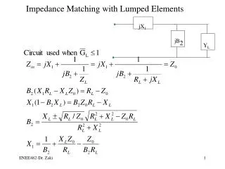
Needle Electrode. Measurement System: Three-electrode method, frequency range 0-200kHz, a thin coaxial needle electrode, large plate (80mm x 160mm) Puls-response method Calculated: extracellular resistance intracellular fluid resistance cell membrane capacitance

E N D
Needle Electrode • Measurement System: • Three-electrode method, • frequency range 0-200kHz, • a thin coaxial needle electrode, • large plate (80mm x 160mm) • Puls-response method • Calculated: • extracellular resistance • intracellular fluid resistance • cell membrane capacitance • Electrical impedance of varius tumors, in vivo. 54 patiens with breast disease. 57 patients with pulmonary disease. “A study of the electrical bio-impedance of tumors”, Morimoto (1993)
Classical four-electrode plunge probe In vivo hepatic tumour and normal tissue conductivity at seven frequencies from 10 Hz to 1 MHz, in rats. Ag-AgCl electrodes The proximal 2 mm of the probe was insulated to reduce errors caused by fluid buildup on the probe surface. “In vivo electrical conductivity of hepatic tumours”, Haemmerich, Staelin, Tsai, Tungjitkusolmun,Mahvi and J. G.Webster (2003) • Planar electrodes • design electrodes as large as possible • use the same geometry for the inner electrodes • - electrode-electrolyte interface impedance as low as possible. • Pt electrodes can be coated with a porous layer of “black platinum” “Design considerations for optimum impedance probes with planar electrodes for bioimpedance measurements.”, Ivorra, A., Aguilo´ , J., Milla´n, J., (2001)
Concentric ring electrode, non-invasive probe The electrode ring system on the tip of the non-invasive probe, The outermost electrode (1) is approximately 10 mm indiameter. The two outer electrodes [(1) and (2)] are sourceelectrodes. The innermost electrode (4) is a current sink.The electrode (3) surrounding the innermost electrode is a guard electrode, which reduces surface currents. Microinvasive probe, voltage is applied at the beams marked (a) and (b), and the current is detected in beam (c) Area of the electrode is 5 x 5 mm2. ”Micromachined electrodes for biopotential measurements” Griss,Enoksson,Tolvanen-Laakso,Meriläinen,Ollmar (2001)
Electrode array Detecting probe: matrix of 8x8 pointed, gold-coated electrodes (4mm x 4 mm) Voltage source: a hand held cylinder (metallic clip) Trans-impedance measurement technique: while the evoked current under a particular electrode was recorded, the other electrodes were kept at ground potenttial. Electrical impedance scanning (EIS), 100 Hz – 100kHz. GN= average of the normalized conductivity “Electrical impedance scanning: a new approach to skin cancer diagnosis”, Glickman (2003)
Parallel plate four-electrode probe Ag-AgCl electrodes 31 logarithmically distributed frequencies, 1 kHz- 1MHz Results show that Electrical impedance spectroscopy (EIS) is able to repeatedly detect small tumors (<3 mm) and tumorassociated changes, whereas CT and US were not routinely capable of detecting pathological developments on this scale. [Intramuscular carcinomas] “Feasibility studies of electrical impedance spectroscopy for early tumor detection in rats”, Skourou et al (2004) “Non-invasive assessement of radiation injury with electrical impedance spectroscopy”, Osterman et al (2004)


![G5 - ELECTRICAL PRINCIPLES [3 exam questions - 3 groups]](https://cdn0.slideserve.com/382273/g5-electrical-principles-3-exam-questions-3-groups-dt.jpg)