Download
1 / 45
570 likes | 1.47k Vues
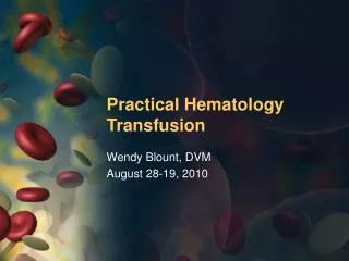
Practical Hematology Blood Loss Anemia. Wendy Blount, DVM August 28-19, 2010. Course Materials. Course Materials. http://www.wendyblount.com Click on “Presentation Notes” Select this course Download materials. Practical Hematology. Determining the cause of anemia

E N D
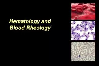
Practical HematologyBlood Loss Anemia Wendy Blount, DVM August 28-19, 2010
Course Materials • http://www.wendyblount.com • Click on “Presentation Notes” • Select this course • Download materials
Practical Hematology • Determining the cause of anemia • Treating regenerative anemias • Blood loss • Hemolysis • Treating non-regenerative anemias • Blood & plasma transfusions in general practice • Determining the causing of coagulopathies • Treating coagulopathies in general practice • Finding the source of leukocytosis • Bone marrow sampling
Diagnosis • “Anemia” is not a diagnosis • It’s a symptom • Treating anemia without knowing the diagnosis doesn’t often work out very well What is the most common treatment for anemia? • Very few anemias require treatment with iron • Iron supplementation will significantly help very few anemias • Contraindicated for anemia of chronic inflammatory disease
Diagnosis When is anemia significant? • Cats – PCV persistently <20-25% • Dogs – PCV persistently <30-35% Symptoms secondary to anemia IOW – when to run a CBC • Reduced oxygen carrying capacity • Tachypnea, dyspnea, syncope • hypoxia without cyanosis • Pallor • Reduced blood volume • Weak peripheral pulses • Pallor, slow CRT (Capillary Refill Time) Related to underlying disease
Diagnosis 2 parts of a CBC • Automated count • Should be run within 3 hours • Blood smear examination • Do on all CBCs with significant abnormalities • Prepare smears within 30 minutes • RBC and WBC morphology • Hemoparasites • Inclusions – Dohle bodies • Differentiate WBC cell lines count can not • Sometimes there are cells that the counter can not identify
Diagnosis Severity of Symptoms • Rapidity of onset • Severity of Anemia • Degree of physical activity (cats) • Concurrent disease affecting respiratory exchange • Respiratory disease • Cardiovascular disease Pseudoanemia • Mild decrease in PCV due to plasma volume expansion, RBC normal • Congestive heart failure, pregnancy, glucocorticoid therapy, IV fluid therapy
Diagnosis The First Question • Is the anemia regenerative? • IOW – is the body losing RBCs or not making them or both? • At maximum stimulation, the bone marrow can make RBCs at 50x the usual rate • It takes at least a few days and up to a week for this to fully kick in • An acute regenerative anemia can look non-regenerative during the first week
Assessing the Regenerative Response Reticulocytes • RNA to make Hb retained for a few days after the nRBC extrudes its nucleus • Mix EDTA blood with stain 1:1 • New methylene blue • Brilliant cresyl blue • Incubate 15 minutes • Count 500-1000 RBC • Report % retics of RBC counted
Assessing the Regenerative Response Reticulocytes • Count only aggregates, not punctates in cats • Punctates have up to 10 blue dots that do not coalesce
Assessing the Regenerative Response Percent Reticulocytes • Non-anemic animals <0.4% retics • >1% usually a regenerative response • This method is not as reliable as… Absolute Reticulocyte Count • RBC/ul x % retics = ARC • Non-anemic animals <40,000/ul • >80,000/ul is a moderately regenerative response • Automated counts are not always reliable • This is the preferred single index for assessing regenerative response
Assessing the Regenerative Response Corrected Percent Reticulocytes • If you don’t know the RBC and can not calculate absolute retics, you can still correct retic % for anemia % retics x patient PCV normal PCV Cat normal PCV = 37%, Dog normal PCV = 45% • Normal animals <0.4% corrected retic % • >1% is a regenerative response
Assessing the Regenerative Response If you can’t calculate an absolute retic count, then corrected retic % (CRP) is second best Reticulocyte Production Index (RPI) • No longer used very much Increased RDW (red cell distribution width) • Objective measure of anisocytosis • If increased, you have one of the following: • Normal + large RBC – regenerative • Small + normal RBC – developing IDA • All 3 cell sizes – chronic blood loss
Assessing the Regenerative Response Increased MCV (mean corpuscular volume) • Often increased with regeneration • Can also be increased with RBC maturation arrest • FeLV • marrow dysplasia • folate deficiency • Phenobarbital therapy • Nonpathogenic congenital disorder • Poodles
Assessing the Regenerative Response nRBCs – normoblasts, metarubricytes • Increased with: • Regenerative anemia • Splenic disease • Bone marrow disease • Iron deficiency anemia • Howell-Jolly Bodies are end stage RBC
Assessing the Regenerative Response RBC morphology – signs of regeneration • Anisocytosis – variation in RBC size • Polychromasia – blue-gray big RBCs • >1/HPF (oil) indicates inc retics
Regenerative Anemia So you know your anemia is Regenerative… Now What???
Regenerative Anemia Degree of Regeneration Acute Blood Loss – non-regenerative, then moderately regenerative 3-7 days later Chronic Blood Loss – marked regeneration Hemolysis – moderate to marked regeneration
Regenerative Anemia RBC morphology • Hypochromasia • Chronic blood loss • Spherocytes • Intravascular hemolysis • IMHA – immune mediated hemolytic anemia • Schistocytes • DIC • Liver or Splenic Disease • Microangiopathy • Hemangiosarcoma • Caval syndrome
Regenerative Anemia RBC morphology An abnormality should be present in nearly every field to be considered significant Senescent cells can display any morphologic abnormality Poikilocytosis = any abnormal RBC cell shape
Regenerative Anemia • Hemolysis • IV hemolysis • Vascular hemolysis • Extravascular hemolysis • Blood Loss • External • Internal • GI/urinary
Blood Loss Anemia • Localized bleeding • Trauma/surgery • Neoplasia or infiltrative disease • Skin - fleas • Tendency for generalized bleeding • coagulopathy
Blood Loss Anemia • Acute Blood Loss • Trauma/surgery • Neoplasia • Bleeding GI ulcer • Abdominal cavity bleeding • Chronic Blood Loss • Fleas or intestinal parasites • GI or urinary tract bleeding • Erosion of external artery • Vasculitis – epistaxis
Acute Blood Loss • Total blood volume • 8-10% of body weight in dogs • 6-8% of body weight in cats <20% blood loss is well tolerated • <8-10 ml/lb in dogs • <6-8 ml/lb in cats • 30-40% blood loss • Hypotension and shock • Weak pulses, cold extremities • Laterally recumbent • 50% blood loss • Can be fatal if over less than 2-3 hours
Acute Blood Loss Response to Acute Blood Loss • Within a few hours • EPO levels rise • Platelets drop no lower than 60,000/ul • Stress leukogram is possible • Within 2-3 days • Bone marrow response begins • Restoration of plasma volume • Following PCV can grossly underestimate acute blood loss
Acute Blood Loss Response to Acute Blood Loss • Maximum regenerative response within 7 days • Corrected retic % can be 3-7% • Absolute retics >100,000/ul • In cats, punctate retics may remain elevated for weeks • May have rebound thrombocytosis • Recovery within 1-2 weeks HALLMARK OF ACUTE EXTERNAL BLOOD LOSS (triad) • Anemia • Hypoproteinemia – albumin and globulin • Reticulocytosis
Treating Acute Blood Loss Stop the Bleeding Replace fluid loss Oxygen support Treat underlying disorder
Treating Acute Blood Loss Stop the Bleeding • Assess coagulation status • External arterial bleeder • Temporary • Cautery - silver nitrate, Kwik Stop, electrocautery • Epinephrine • Permanent • Excise abnormal tissue for biopsy • Reveal normal artery and ligate
Treating Acute Blood Loss Stop the Bleeding • Abdominal bleeder • exploratory surgery as soon as vascular volume and oxygen carrying capacity restored • GI bleeder • Sucralfate PO – 1-3g in a slurry • Barium PO – 3-5 ml/lb • Endoscopic cautery • surgery
Treating Acute Blood Loss Replace fluid loss • crystalloids • 10 ml/lb bolus and then reassess • 1-2 ml/lb/hr when hypovolemia replaced • Colloids • Hetastarch • 10 ml/kg over 5-15 minutes • repeat once if needed • Oxyglobin • 3-5 ml/kg added to fluids running at 0.5-2ml/lb/hr • Or 10 ml/kg/hr for up to 3 hours • If IV access is difficult, try intraosseous
Treating Acute Blood Loss Oxygen support • Transfusion – RBC or whole blood • Oxyglobin • Oxygen – nasal, flow-by, mask, intubate Treat underlying disorder
Treating Acute Blood Loss Transfusion • PCV threshold higher for acute blood loss • 20-25% with signs of hypoxia • Or if going to surgery • Improves oxygen carrying capacity • May improve hemostasis • Normally, transfusion of 10 ml/lb whole blood is given over a minimum of 2 hours • Pretreat with dexamethasone • Give as fast as is tolerated • Collect blood from the abdomen, pass through filter and re-administer (use anticoagulant) • No limitation on administration rate
Chronic Blood Loss CHRONIC EXTERNAL BLOOD LOSS IS THE MOST COMMON CAUSE OF IRON DEFICIENCY ANEMIA IN DOGS AND CATS • Also CRF (chronic renal failure) • Increased gastrin causes GI ulceration • Chronic blood loss is usually markedly regenerative • Increased retics, RDW, anisocytosis • Retics may be >500,000/ul or 10%+ corrected • Polychromasia less pronounced • Only becomes non-regenerative if very chronic • Absent iron stores in issues • liver, spleen and marrow • ferritin - soluble iron stores • Hemosiderin - insoluble iron stores
Chronic Blood Loss • Low serum iron - <60 ug/dl • Low transferrin saturation - <20% • Transferrin is serum protein that transports iron • Normally 20-60% saturated • Determined by measuring UIBC – unbound iron binding capacity, which is increased • Increased TIBC (iron binding capacity) • Increased transferrin
Chronic Blood Loss • Low Hb and HCT
Chronic Blood Loss • Low Hb and HCT • Blood smear • Hypochromasia – pale RBC • Low MCHC • Microcytosis • low MCV – small RBC and leptocytes • RBC become stiffer & more susceptible to lysis • Thrombocytosis • May exceed 1,000,000/ul • Mechanism unknown • Platelets >1 million warrants search for blood loss, if pet is not splenectomized • Low globulins and albumin
Chronic Blood Loss Causes of chronic blood loss and IDA • GI hemorrhage – MOST COMMON • Including inflammatory bowel disease • Both iron malabsorption and bleeding • Ulcer or aneurysm • Neoplasia • Liver disease – coagulopathy and ulcers • Parasitism • Fleas • hookworms • Rarely whipworms • Chronic externally bleeding neoplasia Iron supplementation is rarely needed unless there is chronic external blood loss or CRF
Chronic Blood Loss Clinical Signs • Onset insidious - develops over weeks • Patients may seem quite well for their severe anemia (<15-20%) • Sudden death can occur, when oxygen demands exceed oxygen carrying capacity • Most common presenting signs • Pallor • exercise intolerance – syncope • pica – eating dirt, rocks, etc. • Melena is not always obvious when there is significant chronic GI bleeding • Bleeding can be intermittent • Fecal cytology to look for RBC can help
Chronic Blood Loss Clinical Signs • Decreased blood viscosity • Bounding pulses • Physiologic murmur • Gallop rhythm • Increased blood volume • Cardiac hypertrophy and dilation • congestive heart failure • Depletion of iron from body tissues • Muscle weakness • Abnormal behavior • Dry brittle Skin and nails, hair loss, abnormally shaped nails
Treating Chronic Blood Loss Correct Anemia - Transfusion Treat underlying disorder Correct Iron Deficiency
Treating Chronic Blood Loss Correct Anemia - Transfusion • Anemia severe enough to cause clinical signs (PCV <15-20%) • Or preparing for corrective surgery • Conservative transfusion volume to avoid precipitating CHF • Volume overload more of a problem in cats than in dogs • Use packed cells • Correction of anemia results in resolution of cardiomegaly within several weeks
Treating Chronic Blood Loss Treat Underlying Disorder • Deworm/deflea after patient is stabilized • If GI Bleeding confirmed • Abdominal US • Endoscopy • Exploratory Laparotomy • Confirm blood loss has resolved by monitoring reticulocyte count • < 40,0000/ul • Retics more sensitive than anemia for chronic blood loss
Treating Chronic Blood Loss Correct Iron Deficiency • Ferrous sulfate 5 mg/lb/day PO • Give with a meal • Continue for weeks to months • Serology to confirm iron stores are replete • TIBC – falls back to normal • Transferrin – 20-60% saturated • Iron – 60-230 ug/dl