Practical Hematology
Various other dog breeds (poodles) DNA tests for many breeds. PK enzyme activity test for ... Clear areas in cat RBC are most likely oxidized hemoglobin ...

Practical Hematology
E N D
Presentation Transcript
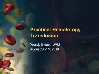
Slide 1:Practical Hematology Hemolytic Anemia
Wendy Blount, DVM August 28-19, 2010
Slide 2:Practical Hematology
Determining the cause of anemia Treating regenerative anemias Blood loss Hemolysis Treating non-regenerative anemias Blood & plasma transfusions in general practice Determining the causing of coagulopathies Treating coagulopathies in general practice Finding the source of leukocytosis Bone marrow sampling
Slide 3:Hemolysis
Normal lifespan of the RBC 100-120 days in dogs 70-80 days in cats Causes of shortened RBC lifespan Premature RBC removal Extravascular hemolysis In the liver, spleen & bone marrow May be triggered by antiRBC Ab Intravascular RBC destruction May be triggered by antiRBC Ab Or complement membrane permeability changes Enzyme deficiency of malfunction
Slide 4:Hemolysis
Clin Path Changes with EV hemolysis, in addition to low PCV Increased serum bilirubin yellow to orange serum Bilirubinuria yellow-orange urine Small amounts bilirubin made by normal canine kidneys Always pathologic in the cat
Slide 5:Hemolysis
Clin Path Changes with IV hemolysis Increased serum Hb amber to red serum Increased serum bilirubin Yellow to orange serum Depleted plasma haptoglobin Hb breaks into 2 dimers that bind to plasma haptoglobin Hemoglobinuria red-brown urine Distinguish from hematuria Few RBC on sediment Myoglobinuria rare in small animals
Slide 6:Hemolysis
Signs of Hemolysis (vs. Blood Loss) Jaundice Gingiva and sclera first Then skin Pigmenturia Bilirubinuria Hemoglobinuria in dogs Hburia not always present in cats who are hemolyzing
Slide 7:Hemolysis
Signs of Hemolysis (vs. Blood Loss) Autoagglutination Indicates immune mediated hemolytic anemia (IMHA)
Slide 8:Checking for Autoagglutination
Gross autoagglutination 1 drop saline and 1 drop blood on the slide Microscopic AutoAg � wet mount 5:1 saline to blood, coverslip Dilute until you can see RBC with space between them Stacks of poker chips is Rouleaux � dilute more Piles of poker chip winnings (stuck to each other) is AutoAg
Slide 9:Checking for Autoagglutination
Slide 10:Checking for Autoagglutination
3. Microscopic AutoAg � stained smear Look at the feathered edge
Slide 11:Checking for Autoagglutination
4. Saline Wash Blood mixed with saline 3:1 to 5:1 Centrifuge and remove supernatant 1-5 times Then dilute for a microscopic wet mount
Slide 12:Causes of Hemolysis
Inherited RBC defects IMHA Transfusion Reaction Neonatal Isoerythrolysis Infection Mycoplasma haemofelis Cytauxzoon felis Babesia canis Babesia gibsoni Bartonella hensalae Hypophosphatemia
Slide 13:Causes of Hemolysis
Toxicity Methemoglobinemia Heinz body anemia Zinc and copper toxicity Naphthalene Onion, garlic, broccoli Propylene glycol Membrane lipid abnormalities Severe liver disease Microangiopathy Caval syndrome hemangiosarcoma
Slide 14:Inherited Hemolytic Disorders
Hyperkalemia with IV hemolysis Some dog breeds keep NaKATPase in their RBC Normal RBC have low K and high Na RBC of these dogs have the reverse Akita, Shiba Inu, Asian mongrels
Slide 15:Inherited Hemolytic Disorders
Stomatocytosis Overhydrated cup-shaped macrocytes Slit-like central pallor Chondrodystrophic Malamutes Schnauzers
Slide 16:Inherited Hemolytic Disorders
Congenital spherocytosis golden retrievers
Slide 17:Inherited Hemolytic Disorders
Congenital Osmotic Fragility Recurring anemia, splenomegaly, weight loss, lymphocytosis, hyperglobulinemia Abyssinian and Somali cats Also seen in Siamese and DSH Pred and splenectomy and reduce phagocytosis or damaged RBC PFK Deficiency (phosphofructokinase) PFK important to anaerobic glycolysis RBC have no nucleus or mitochondria Hemolytic crises and exertional myopathy English Springer & Cocker Spaniels Dx - enzymatic PFK test or DNA test
Slide 18:Inherited Hemolytic Disorders
PK Deficiency (pyruvate kinase) PCV 10-32% Retics as high as 95% No spherocytes Myelofibrosis and osteosclerosis of bone marrow at 1-3 years of age Die of anemia or liver failure due to hemosiderosis in middle age Splenectomy and prednisone do not help dogs, but help cats Basenji�s, Abyssinian, Somali Various other dog breeds (poodles) DNA tests for many breeds PK enzyme activity test for others
Slide 19:Immune Mediated Hemolytic Anemia
Two kinds of IMHA Auto-immune (anti-RBC or complement mediated) Primary � idiopathic Secondary - Disease process triggers anti-RBC antibodies Allo-immune Anti-RBC are produced by another animal Neonatal isoerythrolysis Transfusion reaction
Slide 20:Immune Mediated Hemolytic Anemia
Causes of secondary autoimmune IMHA Neoplasia Lymphoma, myeloma, others Chronic infection Viral � FeLV, FIV, FIP, URI Bacterial � Lepto, Hemobartonella/Mycoplasma, Bartonella, any chronic abscess Parasitic � Babesia, Leishmania, HWDz, Ehrlichia, Hookworms Drug induced TMPS, cephalosporins, penicillin methimazole Vaccination � within 2-4 weeks Toxicity Bee sting, snake bite
Slide 21:Immune Mediated Hemolytic Anemia
Syndromes associated with primary autoimmune IMHA Automimmune diseases SLE � systemic lupus erythematosis Polyendocrine disorder hypothyroidism Diabetes mellitus Addison�s disease Genetic predisposition American cocker spaniel (33%) English Springer Spaniel Old English sheepdog Irish Setter, Poodle, Dachshund
Slide 22: IMHA is the most common cause of hemolytic anemia in dogs Most common causes of HA in cats: FeLV, Hemobartonella Methimazole Chronic inflammation IMHA + IMT = Evan�s Syndrome
Slide 23:Clinical Signs of IMHA
Vomiting or diarrhea are the most common chief complaints Fever Hepatosplenomegaly, lymphadenopathy Followed by clinical signs of anemia Weakness, lethargy, pallor, etc. Signs of cold agglutination disease Skin necrosis at the extremities Ears, nose, tail tip, nail beds Signs of hemolysis Icterus and pigmenturia Signs of thromboembolic Disease Dyspnea � PTE Swelling of head or limbs (vein thrombosis)
Slide 24:Lab Abnormalities of IMHA
Icterus and pigmenturia - hemolysis Neutrophilia (often >100,000/ul) May have degenerative left shift Bone marrow with both erythroid and myeloid hyperplasia Thrombocytopenia If Evan�s Syndrome (<25,000/ul) Or DIC (any thrombocytopenia) ALT and SAP usually elevated Even prior to corticosteroids Spherocytes on blood smear Abnormalities associated with underlying disease
Slide 25:Spherocytes
Slide 26:Spherocytes
Two-thirds of dogs with IMHA have them in large numbers Small in size hyperchromic No central pallor Can be present in smaller numbers with other causes of hemolytic anemia Hypophosphatemia Zinc toxicity Microangiopathy Heartworm disease hemangiosarcoma Spherocytes are a canine phenomenon
Slide 27:IMHA can Appear Nonregenerative
Acute/peracute onset 1 week needed for regenerative response Antibodies can be directed against RBC precursors in the bone marrow If autoagglutination is present without a regenerative response, do a bone marrow Autoimmune bone marrow disease Bone marrow neoplasia (LSA) Ehrlichia, FeLV These animals must be aggressively transfused
Slide 28:Treating IMHA
Monitoring PCV at least BID at first IMHA can vary from mild to life threatening Platelets SID to QOD Look for ITP and DIC Treat the underlying cause All treated with doxycycline 5-10 mg/kg PO/IV BID x 3 weeks Withdraw any drugs with might be causing IMHA
Slide 29:Treating IMHA
Supportive care Rehydrate and maintain hydration Avoid overzealous IV fluid therapy Transfuse packed cells Or whole blood if DIC Little evidence that transfusion worsens IMHA Alloantibodies don�t trigger autoantibodies Autoagglutination makes reading cross-matches difficult Use universal donor blood DEA 1.1 negative Oxyglobin can decrease need for blood More effective when used earlier
Slide 30:Treating IMHA
Specific Therapy � immunosuppression Corticosteroids Prednisone 1-2 mg/lb/day Dexamethasone 0.15-0.3 mg/lb QOD Azathioprine 1 mg/lb SID x 2 weeks, then QOD Takes 10-14 days to kick in � start early in severe cases Cyclosporine 5 mg/lb/day Titrate dose based on blood levels Increase up to 20 mg/kg/day Steady state within 48 hours Trough target 100-500 ng/ml No bone marrow suppression
Slide 31:Treating IMHA
Specific Therapy � immunosuppression 4. Leflunomide (Arava) 4 mg/kg PO SID Plasma trough 20 ug/ml Expensive Pharmacist may have apoplexy over the proposed dose 5. Danazol 5 mg/lb PO SID Specifically for ITP Expensive Possible hepatotoxicity 6. Cyclophosphamide 200 mg/m2 weekly PO or IV Can divide over 3-4 days Usually given no more than 4 times
Slide 32:Treating IMHA
Specific Therapy � immunosuppression IV Immunoglobulin to block antiRBC Human IVIG 0.5 g/lb x 2 days Short lived response expensive Splenectomy Poor response to drugs Drug side effects unacceptable Spleen is major site of autoantibody production and extravascular hemolysis Spleen histopath may identify underlying disease Disastrous if IMHA is secondary to infectious disease Susceptible to overwhelming infection in the future
Slide 33:Treating IMHA
Continue each drug for at least 2-3 days before increasing dose or adding another drug evidence of response Stabilized or rising PCV Resolution of or less autoagglutination Fewer spherocytes Falling bilirubin Continue immunosuppressive corticosteroids for at least 2 weeks before decreasing 2 mg/lb/day for no longer than 2 weeks Longer can risk GI ulceration If regenerative anemia relapses while still on high dose pred, consider GI bleeding
Slide 34:Treating IMHA
Wean off immunosuppressive drugs over 2-4 months after hemolysis has stopped May take 6-12 months, or never happen Change one drug at a time Not more often than every 2 weeks Not more than 1/3 to � of dose require to control hemolysis Reduce drug only when PCV stable for 2 weeks The faster hemolysis is controlled, the faster the weaning process Wean drugs causing most side effects first
Slide 35:Treating IMHA
Treat sequellae All treated with heparin 50 U/kg SC TID Heparin 200 U/kg SC TID or coumadin if thromboembolic disease develops Plasma 5-10 ml/lb daily for DIC Watch carefully for GI ulceration and treat promptly No drugs are proven to decrease risk of GI ulceration Dog more often die of sequellae of disease or treatment than anemia Jugular catheters can result in thrombosis
Slide 36:Treating IMHA
Treating relapses Wean slowly and carefully to prevent relapse Each episode can be more difficult to treat than the last Taper more gradually with each successive successfully treated relapse Preventing relapse Consider never vaccinating the dog again Prevent relapse of underlying cause
Slide 37:IMHA - Prognosis
Negative prognostic indicators Bilirubin >10 mg/dl � grave prognosis Rapid drop in PCV (10-15% in one day) Non-regenerative response Intravascular hemolysis hemoglubinuria Persistent autoagglutination Thromboembolic complications General condition of the patient Not the first episode (relapse)
Slide 38:Infection Associated Hemolysis
Direct infection of the RBC by organism Mycoplasma/Hemobartonella Cytauxzoon Babesia Indirect hemolysis by inflammatory mediators Systemic inflammation causes shortened RBC lifespan in cats Some bacteria produce hemolysins Lepto, Clostridium, Strep, Staph
Slide 40:Cytauxzoon felis
Carried by ticks Two life forms Schizonts and then merozoites in the tissue macrophages Enlarging tissue histiocytes cause venous obstruction, ischemia and organ failure Merozoites infect the RBC Piroplasms in the RBC Parasitemia is late stage disease Do serial blood smears Death can occur within days Rapid illness progressing to death in one week Fever, anemia, icterus, pigmenturia, dyspnea, hepatomegaly, splenomegaly, lymphadenopathy
Slide 41:Cytauxzoon felis
Used to be considered uniformly fatal Leukopenia with left shift due to tissue necrosis Anemia may appear non-regenerative Not enough time for regenerative response Some cats do survive and carrier state is possible Transfusion or oxyglobin Heparin 100-200 U/kg SC TID Diminazene (not approved in the US) Imidocarb 2-5 mg/kg IM, repeat in 4-7 days Atovaquone and azithromycin Some cats survive with supportive care PCR is available
Slide 42:Cytauxzoon felis
Slide 44:Mycoplasma-Hemobartonella
Large (more pathogenic) and small types � hemoplasma mollicutes Mycoplasma haemofelis Mycoplasma haemominutum Mycoplasma haemocanis � rare cause of disease Opportunistic 50% of affected cats are FeLV positive PCR is available and a good test, but presence of organism does not always result in disease Make smears immediately after blood collection Organisms detach with time
Slide 45:Mycoplasma-Hemobartonella
Treatment Doxycycline 5 mg/kg PO BID x 3 weeks Enrofloxacin 5 mg/kg PO SID x 3 weeks Both in refractory cases Prednisone 1-2 mg/lb/day x 2 weeks, then taper for secondary IMHA Hemobartonella vs Cytauxzoon Hemobartonella is epicellular Cytauxzoon is intracellular Cytauxzoon in macrophages Hemobart � marked regeneration Cytauxzoon � organ failure
Slide 47:Babesia
Carried by ticks - Babesia canis, Babesia gibsoni Endemic in greyhound and pitbull kennels Puppies more susceptible Transmitted by transfusion Severe hemolytic anemia, fever, shock and multiple organ failure Thrombocytopenia and leukocytosis Lymphadenopathy, splenomegaly Secondary IMHA causes more hemolysis than the organism does directly IFA and ELISA are available Imidocarb 3.5 mg/lb IM Prednisone 1-2 mg/lb/day x 2 weeks, then taper for secondary IMHA
Slide 48:Methemoglobinemia
Heme is oxidized from ferrous to ferric so that it can�t bind and transport oxygen Cats are especially susceptible more sulfhydryl groups on their hemoglobin Also they are glucuronyl transferase is deficient Drugs Benzocaine containing skin products and sprays Acetaminophen Phenazopyridine (urinary tract analgesic) Dyspnea, cyanosis, ataxia, coma, death Facial edema and GI signs with Tyelonol Diagnose with �spot test� Put drop of affected EDTA blood on white paper towel next to control blood Control will turn red, metHb blood stays brown
Slide 50:Heinz Body-Membrane Injury Anemia
Denatured hemoglobin forms Heinz bodies Cats are especially susceptible more sulfhydryl groups on their hemoglobin to oxidize 10% Heinz bodies can be normal in cats Heinz bodies in dogs usually pathologic
Slide 51:Heinz Body-Membrane Injury Anemia
Foods onion, garlic Broccoli propylene glycol � semimoist foods Drugs/Supplements Acetaminophen Methionine Phenothiazines Phenacitin VitaminK3 Propofol � no maintenance drips, not on successive days
Slide 52:Heinz Body-Membrane Injury Anemia
Chemicals naphthalene � moth balls and toilet cleaner Zinc hardware and pennies (since 1983) Serum zinc > 5 ppm (plastic tubes) Copper, phenols Metabolic Disease Diabetic ketoacidosis
Slide 53:Heinz Body-Membrane Injury Anemia
Treatment Remove the oxidative agent antioxidants Methylene blue 0.4 mg/lb slowly IV N-acetylcysteine 64 mg/lb initial dose then 32 mg/lb TID x 7 treatments (PO or IV) Mucomyst is preferred for Tylenol toxicity Vitamin C, vitamin E Transfusion with packed cells Oxygen not all that helpful without transfusion
Blister cells keratocytes Eccentrocytes Clear areas in cat RBC are most likely oxidized hemoglobinSlide 57:Hypophosphatemia
Hemolysis can occur when phosphorus falls below 2.5 mg/dL Likely to occur when < 1.5 mg/dL Causes of severe hypophosphatemia DKA � diabetic ketoacidosis Hepatic lipidosis Refeeding syndrome Phosphate binder overdose (AlOH) Supplement phosphorus when feeding anorectic cats, or treating DKA IV � potassium phosphates (chart) Watch for hypocalcemia PO when eating and stable � most foods contain plenty of phosphorus Dilute tube feeding diets with skim milk
Slide 59:Microangiopathy
Schistocytes Triangular or helmet shaped RBC fragments Destruction of RBC as they move through damaged blood vessels Endocarditis Hemangiosarcoma Caval Syndrome � Heartworm Disease Thrombosed IV catheter Vasculitis Hemolytic-uremic syndrome DIC Schistocytes are also seen with osmotic fragility Liver disease, iron deficiency, water intoxication, congenital, zinc toxicity
Slide 60:Work-Up for Hemolytic Anemia
CBC with reticulocyte count General Health profile with electrolytes Urinalysis with sediment FeLV/FIV for cats, occult HWTest for dogs Fecal Check for autoagglutination Blood smear cytology Chest and abdominal x-rays Abdominal US Aspirates of spleen, liver, lymph nodes if enlarged Coag panel if thrombocytopenic or critically ill
Slide 61:Work-Up for Hemolytic Anemia
Secondary tests � as indicated by symptoms DNA or enzyme activity tests if congenital hemolytic anemia is suspected Tick panel Bartonella PCR/culture Lepto titers Thyroid panel ANA � support diagnosis of IMHA Antiplatelet antibodies if thrombocytopenic COOMBS TEST IS NOT ON THIS LIST