Download
1 / 20
200 likes | 213 Vues
This study aims to develop a teledermatology application that includes a database and software for comparing and classifying various skin diseases. The application will assist dermatologists in disease diagnosis and prognosis, ultimately improving patient care.

E N D
A novel approach for diagnosis and prognosis of skin diseases using image analysis Guide: Dr. ManjunathShenoyProfessor and H.O.D. Dept. of Dermatology Yenepoya Medical College Yenepoya UniversityMangalore Student: Parameshwar R. HegdeJunior Research Fellow Dept. of Dermatology and Yenepoya Research Centre Yenepoya UniversityMangalore
Contents • Background • Introduction • Literature survey • Rational of study • Aim and objectives • Relevance of study • Materials • Methods • Timeline • Budget • Social relevance
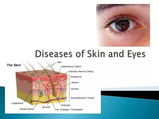
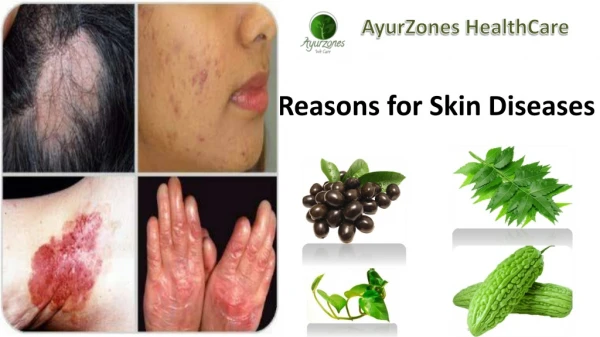
Background for study • Skin diseases are important health problems which also impair the quality of life (Kurpiewska et al., 2011) • The lesions seen in skin diseases may vary in severity from minor localized patches to complete body coverage (Ho, 2010) • Diagnosis mainly depends on the experience of the clinician • Human judgement may have discrepancies in quantification of information gathering, accuracy, reproducibility of data (Broek et al., 2011) • Biopsy for histopathology is, invasive test and also time consuming (Manocha et al., 2011)
Introduction • Image analysis is the extraction of relevant information from images using the digital image processing techniques (Gonzalez and Woods, 2002) • It is very closely related to the medical imaging field, medical image analysis focuses on the computational analysis • The methods can be grouped into several categories: image acquisition, image segmentation, feature extraction (Gonzalez and Woods, 2002) • Machine learning draws results from artificial intelligence, probability and statistics, control theory (Mitchell, 1997)
Table 1. International scenario of the study. Literature survey
Table 2. National scenario of the study. Literature survey
Rationale of study • The available information related to current work is scarce • Currently 15 working online databases are available worldwide (Dermnet, 1998; Hardin.MD, 2010) and no authentic online databases are available from India • Existing databases has limitations like, updation, diseased image metadata, user registration, prognosis tool • Earlier disease classification tools, classified specific disease from normal skin and other 6 related skin diseases
Relevance of study • Teledermatology services will be easily instituted for patients with skin ailments • Assist the dermatologists in disease prognosis
Aim of study • To develop a teledermatology application including database and software to compare and classify various skin diseases
Objectives of study • To collect the diseased image and create a database • Use the database for teledermatologyapplication • To develop a classification tool for skin diseases
Materials • A camera to collect the images (Canon 1200D with 18-55 and 50mm lens) • A high performance computer (1TB HDD, 8GB RAM, i7 processor, 2GB graphic card, 18.5” TFT) for programming • MATLAB tool to develop the machine learning based diagnosis software • Server to host teledermatology application
Methods • Data (image) collection • White or dark background using digital camera • Sample size – 1000 images (Arfin et al., 2012, Yusof et al., 2011) • Sample – Normal skin, Diseased lesion (Plaque psoriasis, Chronic eczema, Lichen planus, Pityriasisrosea) • Sample source – Department of skin and VD, Yenepoya medical college, YenepoyaUniversity • Naming each image with a unique code and create a database in MS Excel
Methods • Teledermatology application • Creating application backend by collected information • Backend – MySQL • Developing graphical interface for user • Frontend – PHP, Java Scripts • Diagnosis software • Pre-processing – Removing unwanted information from image • Hair removing, cropping • Feature extraction – Extracting useful regions from image • Color and texture features
Methods • Classification – Making a decision based on extracted feature • Statistical analysis • Sensitivity, specificity and accuracy
Time line Figure 1. Work plan (Gantt chart) of the proposed study.
References • Arifin, ML., Kibria, MG., Firoze, A., Amin, MA., Yan, H. (2012). Dermatological disease diagnosis using color skin images. International Conference on Machine Learning and Cybernetics, Xain. p. 1675-1680. • Broek., Harris, N., Henkens, M., palma, PP., Szumilin, E. (2013). Introduction. In: Giouzard V, editor. Clinical guidelines: Diagnosis and treatment manual. Medicines Sans Frontieres. pp. 10-12. • Cheerla, N., Frazeir, D. (2014). Automatic melanoma detection using multi-stage neural networks. International Journal of Innovative Research in Science, Engineering and Technology 3:9164-9183. • New Zealand Dermatological Society (1998). Dermnet, viewed 14 June 2016,http://www.dermnet.com/dermatology-pictures-skin-disease-pictures/. • Egmal, M. (2013). Automatic skin cancer images classification. International Journal of Advanced Computer Science and Applications 4:287-294. • Giotis, I., Molders, N., Land, S., Biehl, M., Jonkman, MF., Petlov, N. (2015). MED-NODE: A computer assisted melanoma diagnosis system using non-dermoscopic images. Expert Systems with Application 42:6578-6585.
References • Gonzalez, RC., Woods, RE. (2002). Introduction. Digital image processing. Pearson Educational International. pp. 24-29. • University of Iowa (2010). Hardin.MD, viewed 14 June 2016, http://hardinmd.lib.uiowa.edu/index.html. • Hashim, H., Ramli, S., Wahid, N., Hassan, N. (2013). Recognition of psoriasis features via Daubechies D8 wavelet technique. International journal on smart sensing and intelligent systems 6. • Ho, KM. (2010). Psoriasis. Medical bulletin 15:10-14. • Jaleel, JA., Salim, S., Aswin, RB. (2012). Artificial neural network based detection of skin cancer. International Journal of Advanced Research in Electrical, Electronics and Instrumentation Engineering 3:200-205. • Kavitha, J., Saravanan, D. (2014). Localizing 2-D digital skin images using savc algorithm. International Journal of Innovative Research in Science, Engineering and Technology 3. • Kumar, KS., Reddy, BE. (2014). Wound image analysis using contour evolution. I.J. Image, Graphics and Signal Processing 6:36-42.
References • Kurpiewska, J., Liwkowicz, J., Benczek, K. (2011). A survey of work-related skin diseases in different occupations in Poland. International Journal of Occupational Safety and Ergonomics 17:207-214. • Mitchell, T. (1997). Introduction. In: Book news, editors. Machine Learning. Morgan Kaufmann. pp. 1-6. • Manocha, D., Bansal, N., Farah, RS. (2011). Types and selection criteria for various skin biopsy procedures. In: Dr. Khopkar U, editors. Skin biopsy – perspectives. INTECH Open Access Publisher. pp. 35-39. • Shrivastava, VK., Londhe, ND., Sonawane, RS., Suri, JS. (2015). Reliable and accurate psoriasis disease classification in dermatology images using comprehensive feature space in machine learning paradigm. Expert Systems with Applications 42:6184-6195. • Suvarna, M., Sivakumar., Kumar, K., Niranjan, UC. (2013). Diagnosis of burn images using template matching, k-nearest neighbor and artificial neural network. International Journal of Image Processing 2:109-226. • Yusof, YW., Hashim, H., Sulaiman, KA. (2011). Plaque lesion classification fuzzy model based on various color models. The Third International Conference on Advances in System Simulation, Barcelona. p. 88-93.