Download
1 / 5
50 likes | 66 Vues
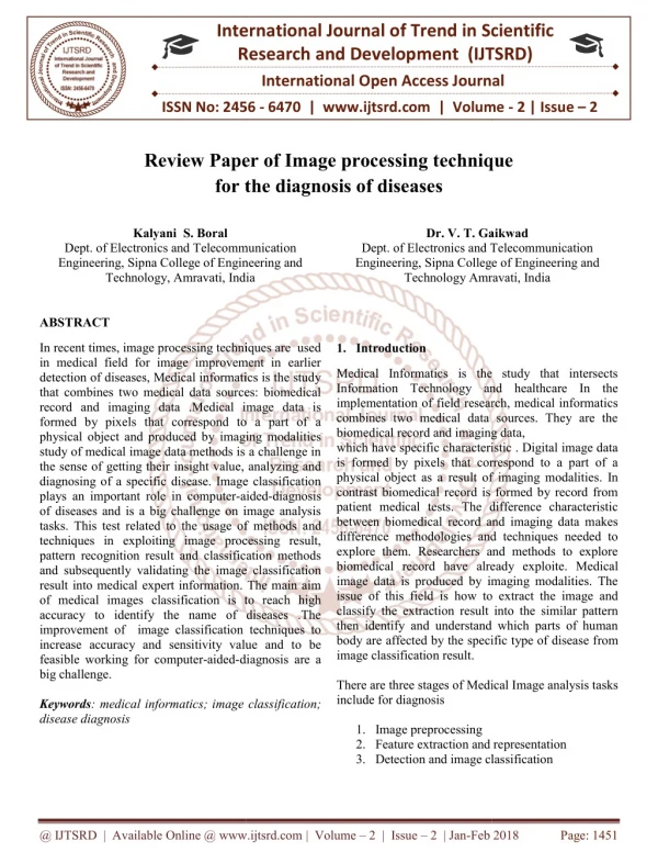
In recent times, image processing techniques are used in medical field for image improvement in earlier detection of diseases, Medical informatics is the study that combines two medical data sources biomedical record and imaging data .Medical image data is formed by pixels that correspond to a part of a physical object and produced by imaging modalities study of medical image data methods is a challenge in the sense of getting their insight value, analyzing and diagnosing of a specific disease. Image classification plays an important role in computer aided diagnosis of diseases and is a big challenge on image analysis tasks. This test related to the usage of methods and techniques in exploiting image processing result, pattern recognition result and classification methods and subsequently validating the image classification result into medical expert information. The main aim of medical images classification is to reach high accuracy to identify the name of diseases .The improvement of image classification techniques to increase accuracy and sensitivity value and to be feasible working for computer aided diagnosis are a big challenge. Kalyani S. Boral | Dr. V. T. Gaikwad "Review Paper of Image processing technique for the diagnosis of diseases" Published in International Journal of Trend in Scientific Research and Development (ijtsrd), ISSN: 2456-6470, Volume-2 | Issue-2 , February 2018, URL: https://www.ijtsrd.com/papers/ijtsrd10703.pdf Paper URL: http://www.ijtsrd.com/engineering/electronics-and-communication-engineering/10703/review-paper-of-image-processing-technique-for-the-diagnosis-of-diseases/kalyani--s-boral<br>

E N D
International Research Research and Development (IJTSRD) International Open Access Journal Review Paper of Image processing technique for the diagnosis of diseases International Journal of Trend in Scientific Scientific (IJTSRD) International Open Access Journal ISSN No: 2456 ISSN No: 2456 - 6470 | www.ijtsrd.com | Volume 6470 | www.ijtsrd.com | Volume - 2 | Issue – 2 Review Paper of Image processing technique for the Kalyani S. Boral Dr. V. T. Gaikwad T. Gaikwad Dept. of Electronics and Telecommunication Engineering, Sipna College of Engineering and Technology, Amravati, India echnology, Amravati, India mmunication ngineering and Dept. of Electronics and Telecommunication Engineering, Sipna College of Engineering and Dept. of Electronics and Telecommunication Engineering, Sipna College Technology Amravati, India echnology Amravati, India ABSTRACT In recent times, image processing techniques are used in medical field for image improvement in earlier detection of diseases, Medical informatics is the study that combines two medical data sources: biomedical record and imaging data .Medical image data is formed by pixels that correspond to a part of a physical object and produced by imaging modalities study of medical image data methods is a challenge in the sense of getting their insight value, analyzing and diagnosing of a specific disease. Image classification plays an important role in computer-aided of diseases and is a big challenge on image analysis tasks. This test related to the usage of methods a techniques in exploiting image processing result, pattern recognition result and classification methods and subsequently validating the image classification result into medical expert information. The main aim of medical images classification is to reac accuracy to identify the name of diseases .The improvement of image classification techniques to increase accuracy and sensitivity value and to be feasible working for computer-aided-diagnosis are a big challenge. Keywords: medical informatics; image classification; disease diagnosis In recent times, image processing techniques are used in medical field for image improvement in earlier detection of diseases, Medical informatics is the study combines two medical data sources: biomedical record and imaging data .Medical image data is formed by pixels that correspond to a part of a physical object and produced by imaging modalities study of medical image data methods is a challenge in of getting their insight value, analyzing and diagnosing of a specific disease. Image classification 1.Introduction Medical Informatics is the study that intersects Information Technology and healthcare In the implementation of field research, medical informatics combines two medical data sources. They are the biomedical record and imaging data, racteristic . Digital image data is formed by pixels that correspond to a part of a a result of imaging modalities. In contrast biomedical record is formed by record from patient medical tests. The difference characteristic dical record and imaging data makes difference methodologies and techniques needed to explore them. Researchers and methods to explore ecord have already exploite. Medical image data is produced by imaging modalities. The how to extract the image and classify the extraction result into the similar pattern then identify and understand which parts of human body are affected by the specific type of disease from Medical Informatics is the study that intersects Information Technology and healthcare In the implementation of field research, medical informatics combines two medical data sources. They are the biomedical record and imaging data, which have specific characteristic . Digital image data is formed by pixels that correspond to a part of a physical object as a result of imaging modalities contrast biomedical record is formed by record from patient medical tests. The difference characteristic between biomedical record and imaging data makes difference methodologies and techniques needed to explore them. Researchers and methods to explore biomedical record have already exploite image data is produced by imaging modalities. The issue of this field is how to extract the image and classify the extraction result into the similar pattern then identify and understand which parts of human body are affected by the specific type of disease f image classification result. There are three stages of Medical I include for diagnosis 1.Image preprocessing 2.Feature extraction and representation 3.Detection and image classification aided-diagnosis of diseases and is a big challenge on image analysis tasks. This test related to the usage of methods and techniques in exploiting image processing result, pattern recognition result and classification methods and subsequently validating the image classification result into medical expert information. The main aim of medical images classification is to reach high accuracy to identify the name of diseases .The improvement of image classification techniques to increase accuracy and sensitivity value and to be diagnosis are a There are three stages of Medical Image analysis tasks age classification; Feature extraction and representation Detection and image classification @ IJTSRD | Available Online @ www.ijtsrd.com @ IJTSRD | Available Online @ www.ijtsrd.com | Volume – 2 | Issue – 2 | Jan-Feb 2018 Feb 2018 Page: 1451
International Journal of Trend in Scientific Research and Development (IJTSRD) ISSN: 2456-6470 applications in image processing. Color, texture edges, morphology are the features which are used in disease detection. Monica jhuria et al took color, morphology, texture as feature for the disease detection. It is found that morphological result gives more result than any other features. Texture shows how the color is distributed in the image, hardness of the image. In Feature Extraction here we are using algorithm, which first extract the feature of image and after the whole implementation of process it stores the new image on server and extract features from transformed server. The transformation functions transformation is composed of a sequence of low pass and high pass filters, known as filter bank. The first step is to denoise the image; by using filter the noise is removed. The next step is to normalize the image by using the segmentation by the thresholding then, removal of the infected area named as a region of interest by the morphological operation of dilation and erosion. The next step is boundary detection, to find the infected part by using the edge detection method. Then the values are calculated by using the neural network and then classify whether the input image is infected or not. 2.Literature Review algorithm basis of Figure 1.1 Image processing stages Moreover, important role of image classification. Furthermore medical image classification has three main steps: preprocessing, feature extraction and classification. After preprocessing step then it needs to extract features of interest part from the image for further analysis. The purpose of the pattern classification system is to map the input variables (such as record data or image data) become the output variables (to represent one specific class (with a disease or with no a disease class) . Image classification is a big challenge on image analysis tasks, especially the selection of methods and techniques in exploiting the result of image processing and pattern recognition, classification methods, subsequently validating the image classification result into medical expert knowledge. The main objective of medical images classification is not only to reach high accuracy but also to identify which parts of human body are infected by the disease In the future, an automatic diagnosis technique with image data is needed to be developed for better clinical care. Since image classification research is still an open area and big challenge to validate the image classification result into medical expert knowledge, this paper focuses only on detailed review of mining medical image classification technique with the state-of-the-art that addressing this issue. This paper will not review about feature extraction and feature selection. The aim of the review is to give the reader wide-ranging review of mining medical image classification techniques includes their pros and cons. Feature Extraction is an important part in the disease detection. It plays an important role in identification of an object. Feature extraction is used in many computer-aided-diagnosis needs the Shamsul I. Chowdhury [1] discusses issues related to the analysis and interpretation of medical data in 1994, thus allowing knowledge discovery in medical databases. He also showed that knowledge can also be effectively extracted from a database of patient observations and from interpretation of those observations. The system shows how retrospectively collected data could be utilized for the purpose of knowledge extraction. The main emphasis was to Study the feasibility of the approach exploring a large patient record system. The analysis was carried out to test the hypothesis of a possible causation between hypertension and diabetes. Basically, the medical diagnosis process can be interpreted as a decision making process, during which the physician induces the diagnosis of a new and unknown case from an available set of clinical data and from his/her clinical experience. At the University of Calabria in Italy, the medical decision making process has been computerized, Physicians at the Cosenza General Hospital currently arc using the diagnostic decision support system lo help them with @ IJTSRD | Available Online @ www.ijtsrd.com | Volume – 2 | Issue – 2 | Jan-Feb 2018 Page: 1452
International Journal of Trend in Scientific Research and Development (IJTSRD) ISSN: 2456-6470 the timely identification of breast cancer in patients through The application of a well-defined set of classification data. Dr. Mimmo Conforti presented the system before the ITTS-TTAB’99 audience in 1999 & he explained the architecture horn this particular poiul of view, emphasizing the powerful efficiency and effectiveness of Mathematical approaches as the basic tools for the design of the CAMD or Computer Aided Medical Diagnosis system. Mimmo Cnnforti addresses our attention to cancer early detection on the basis of small arnount of clinical information. Hubert Kordylewski [3] , Daniel Graupe [2] describes the application of a large memory storage and retrieval (LAMSTAR) neural network to Medical diagnosis and medical information retrieval problems in the year 2001. The network also employs features of forgetting and of interpolation and extrapolation, thus being able to handle incomplete data sets. Applications of the network to three specific medical diagnosis problems are described: two from nephrology and one related to an emergency-room drug identification problem.Jenn- Lung Su, Guo-Zhen Wu [6] introduced the database concept has been widely used in medical information system for processing large volumes of data in 2001. Symbolic and numeric data will define the need for new data analysis techniques and tools for knowledge discovery. Three popular algorithms for data mining which includes Bayesian Network (BN), C4.5 in Decision Tree (DT) , and Back Propagation Neural Network (BPN) were evaluated. The result shows that BN had a good presentation in diagnosis ability. to increase classification accuracy in 2007. Early and accurate diagnosing of various diseases has proved to be of vital importance in many health care processes. In recent years intelligent systems have been often used for decision support and classification in many scientific and engineering disciplines including health care. However, in many cases the proposed treatment, prediction or diagnose can differ from one intelligent system to another, similar to the real world where different medical specialists may have different opinions The main aim here is to mimic this real world situation in the manner to merge different opinions generated by different intelligent systems using the self organizing abilities of cellular automata. Michele Berlingerio, Francesco Bonchi, Fosca Giannotti, Franco Turini [4] introduced the concept of Time- Annotated Sequence(TAS) in 2008.Time- annotated sequences(TAS), is a novel mining paradigm that solves this problem. Recently defined in our laboratory together with an efficient algorithm for extracting them, TAS are sequential patterns where each transition between two events is annotated with a typical transition time that is found frequent in the data. The TAS mining paradigm is applied to clinical data regarding a set of patients in the follow-up of a liver transplantation. The aim of the data analysis is that of assessing the extracorporeal photpheresis (ECP) as a therapy to prevent rejection in solid organ transplantation 3.System Architecture A. Block Diagram Programming effectiveness of the B. Description : Figure 2.1 Figure Of Knowledge Discovery Peter Kokol, Petra Povalej, Gregor Stiglic1, Dejan Dinevski [5] elaborates the use of self organization to integrate different specialist’s opinions generated by different intelligent classifier systems with a purpose Diseases are frequent problem to every person and various types of diseases caused by infections are becoming very frequent. We know that all of these diseases are very unsafe, especially if not controlled at an early on stage. infections not only damage the skin. @ IJTSRD | Available Online @ www.ijtsrd.com | Volume – 2 | Issue – 2 | Jan-Feb 2018 Page: 1453
International Journal of Trend in Scientific Research and Development (IJTSRD) ISSN: 2456-6470 It can have a large effect on a person’s daily life, destroy confidence of a person, sling their movement, and turn to depression. Sometimes, many people try to care for these allergies by using their own therapy. However, if these methods are not appropriate for that type of disease then it would make it more dangerous. diseases can simply transfer from human to human so there is a need to control it their initial stage to check it from spreading. This project presents an implementation of a diseases diagnosis system which helps user to detect human diseases and provides medical treatments timely. For this purpose, user will have to upload a disease affected image to our system and give answers to the questions which are asked to user according to the symptoms of the skin. These symptoms are used to recognize the disease and provide a medical treatment. This system works on technologies like image processing and data mining for diseases detection. So the whole project is divided in to below major parts, Image preprocessing, segmentation and feature extraction. Classification model and disease predication. Medical treatment suggestions or advice. The image of disease is taken and various preprocessing techniques are applied onto that image for noise removal and image enhancement. This image is segmented by using a segmentation technique i.e. thresholding segmentation. At last, data mining techniques are used to recognize the disease and to provide recommendations to users. Both image based technique and question naire technique help to increase dependability and performance of the system. Processing step involved following technique : Image preprocessing is to remove noise from the image or other object removal, different preprocessing techniques. Here we are using transformation algorithm. algorithm is used to transform RGB image into grey scale image. In preprocessing involved image enhancement. Image enhancement techniques can be divided into two categories: Spatial domain methods and frequency domain methods. alas, there is no universal theory for determining what “good” image enhancement is when it comes to human observation. If it look good, it is good. However, when image enhancement techniques are used as pre- processing tools for other techniques, the quantitative measures can conclude which techniques are most proper. In the image enhancement stage we used the following three techniques: Gabor filter, Auto-enhancement and Fast Fourier transform techniques. Pixel classification or Mosaic is any technique used in editing images or video, whereby an image is blurred by displaying part or all of it at a clearly lower resolution. It is primarily used for censorship. The result is a standard graphics filter, vacant in all but the most basic bitmap graphics editors. In image segmentation, Thresholding is one of the most great apparatus. The segmented image obtained from thresholding has the reward of lesser storage space, fast processing speed and ease in conduct, compare with gray level image which normally contains 256 levels. techniques have drawn a lot of awareness during the past 20 years . Thresholding is a non-linear action that converts a gray-scale image into a binary image where the two levels are assigned to pixels that are below or above the particular threshold value. In this research, Otsu’s method that uses (gray thresh) role to compute total image threshold is used. Otsu’s method is based on threshold selection by statistical criteria. Otsu not essential minimize the weighted sum of within-class variances of the object and background pixels to found an optimum threshold recall that minimization of within-class variances is the same to maximization of between-class difference. This method gives suitable results for bimodal histogram images. Threshold values based on this method will be between 0 and 1, after achieving the threshold value; image will be segmented based on it. Fuzzy logic is an branch of Boolean logic by Lot_ Zadeh in 1965 based on the mathematical theory of fuzzy sets, which is a simplification of the classical set theory. By introducing the notion of degree in the proof of a form, thus enabling a condition to be in a state other than true or false, fuzzy logic provides a very valuable elasticity for reasoning, which makes it possible to take into account inaccuracy and reservations .One advantage of fuzzy logic in order to formalize human reasoning is that the rules are set in normal language. In this detection method fuzzy logic is used for the conclusion. For the accurate conclusion apply the rules that is fuzzy rules. Result analysis and detection of Disease: The detection of the diseases and with the help of disease classify the infected image with the disease matches Therefore, thresholding image processing @ IJTSRD | Available Online @ www.ijtsrd.com | Volume – 2 | Issue – 2 | Jan-Feb 2018 Page: 1454
International Journal of Trend in Scientific Research and Development (IJTSRD) ISSN: 2456-6470 with the given dataset. For the disease detection and classification, we are implementing the deep learning algorithm. algorithm is used to classify the specified image into appropriate disease hence it will be easy to detect the disease and find out the remedy over the disease. C. Algorithm References 1.S . I. Chowdhury.: Statistical Expert Systems - A Special Application areafor Knowledge-based Computer Methodology. Linkoping Studies in Scienceand Technology, 104.,Department of Computer and Information Science, University of Linkoping, Sweden. Thesis No 2.D. Graupe and H. Kordylewski, “A large memory storage and retrieval neural network for adaptive retrieval and diagnosis,” Int. J. Software Eng. Knowledge Eng., vol. 8, no. 1, pp. 115–138, 1998. 1.Input Image 2.Preprocess an Image 3.Classify Image Pixels into background, ROI and Infected Pixels. 4.Mark infected pixels. 5.Measures dimension of infected region i.e. height and width. 6.Perform Image segmentation so as to separate background, ROI and infected region. 7.Add infected region to database with proper naming. 8.Perform result analysis conclusion. 9.Stop 4.Conclusion 3.H. Kordylewski and D. Graupe, “Applications of the LAMSTAR neural network to medical and engineering diagnosis/fault detection,” in Proc7th Artificial Neural Networks in Eng. Conf., St. Louis, MO, 1997. 4.M. Berlingerio, F. B. F. Giannotti, and F. Turini, “Mining clinical data with a temporal dimension: A case study,” in Proc. IEEE Int. Conf. Bioinf Biomed., Nov. 2–4, 2007, pp. 429–436. to get accurate 5.Kokol P, Povalej, P., Lenič, M, Štiglic, G.: Building classifier international conference on cellular automata for research and industry, ACRI 2004, Amsterdam, The Netherlands, October 25-27, 2004. (Lecture notes in computer science, 3305). Berlin: Springer, 2004, pp. 823-830. cellular automata. 6th Medical image classification is an interesting research area, it combines the diagnosis problem and analysis purposes in the medical field. This paper has provided the detailed review of image classification techniques for diagnosis of human body disease include imaging modalities used, each dataset and pros and cons for each technique. For the future work, the improvement of image classification techniques will increase accuracy value and subsequently feasible to be employed for computer-aided-diagnosis, and more robust methods are being developed. 6.G.Z. Wu, “The application of data mining for medical database”, Master Thesis of Department of Biomedical Engineering, University, Taiwan, Chung Li, 2000. Chung Yuan 7.Randolph A Miller, “Medical Diagnostic Decision Support Systems—Past, Present, And Future – A Threaded Bibliography and Brief Commentary”, JAMIA1994;1:8-27 doi:10.1136/jamia.1994.95236141. 5.Advantages 1. To get a disease detection system in which pathology admin will upload patient’s scanned report. 2.To get an expert system for patients. 3.This system can provide quality medical care. 4.The use of this system can free the radiologist from the routine task. 5.It provides more accurate measurements that may lead to more cons 8.Bell, Michael (2010). SOA Modeling Patterns for Service-Oriented Discovery and Analysis. Wiley & Sons. pp. 390. ISBN 978-0470481974. 9.Wasan, S., Bhatnagar, V., Kaur, H.m “The impact of data mining techniques on medical diagnostics,” Data Science Journal 5, 119–126, 2006. @ IJTSRD | Available Online @ www.ijtsrd.com | Volume – 2 | Issue – 2 | Jan-Feb 2018 Page: 1455