Auditory Perception
Auditory Perception. Rob van der Willigen http://www.mbfys.ru.nl/~robvdw/DGCN22/Anatomy_Physiology/DGCN22_2011_Anatomy_Physiology_Part2.ppt. General Outline P4. P4: Auditory Perception. - Cochlear Mechanotransduction. P5: Auditory Perception. - Physiology of the Auditory Nerve.

Auditory Perception
E N D
Presentation Transcript
Auditory Perception Rob van der Willigen http://www.mbfys.ru.nl/~robvdw/DGCN22/Anatomy_Physiology/DGCN22_2011_Anatomy_Physiology_Part2.ppt
General Outline P4 P4: Auditory Perception - Cochlear Mechanotransduction P5: Auditory Perception - Physiology of the Auditory Nerve
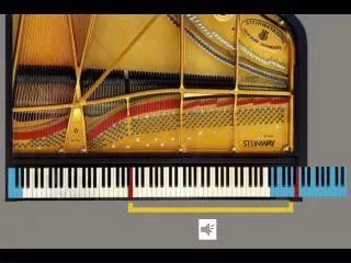
The Problem of Hearing Tonotopie blijft in het auditief systeem tot en met de auditieve hersenschors behouden. “De samenstelling van een geluid uit afzonderlijke tonen is te vergelijken met de manier waarop wit licht in afzonderlijke kleuren uiteenvalt wanneer het door een prisma gaat .” John A.J. van Opstal (Al kijkend hoort men, 2006; p. 8)
The Problem of Hearing Mapping can be an important clue to the function of an area. If neurons are arrayed according to the value of a particular parameter, then that property might be critical in the processing performed by that area. Neurons within a brain area may be organized topographically (or in a map), meaning that neurons that are next to each other represent stimuli with similar properties. Neurons do not need to be arranged topographically along the dimensions of the reference frame that they map, even if its neurons do not form a map of that space.
The Problem of Hearing Problem I: Sound localization can only result from the neural processing of acoustic cues in the tonotopic input! Problem II: How does the auditory system parse the superposition of distinct sounds into the original acoustic input?
Sensory Coding of Sound Outer Hair cells Organ of Corti Inner Hair cell Auditory nerve Basilar Membrane Summary
Sensory Coding and Transduction Mammalian Auditory Pathway Cochlear Mechanotransduction Recapitulation previous lectures
Sensory Coding and Transduction 6 critical steps Recapitulation previous lectures
Sensory Coding and Transduction Peripheral Auditory System The Organ of Corti mediates mechanotransduction: The cochlea is filled with a watery liquid, which moves in response to the vibrations coming from the middle ear via the oval window. As the fluid moves, thousands of hair cells are set in motion, and convert that motion to electrical signals that are communicated via neurotransmitters to many thousands of nerve cells.
Cochlear nonlinearity Active processing of sound CF= 9 kHz OUTPUT Response in dB ~4.5kHz INPUT level (dB SPL) Frequency [kHz] The response of the BM at location most sensitive for ~ 9 KHz tone (CF). The level of the tone varied from 3 to 80 dB SPL (iso-intensity contours). Figure 3. Vertical lines mark the responses to a tone at either 4.5 or 9 kHz. BM input-output function for a tone at CF (~9 kHz, solid line) and a tone one octave below (~4.5 kHz) taken from the iso-intensity contour plot.
Cochlear nonlinearity Active processing of sound A: Tuning characteristic, from the basal turn of the cochlea. Data is shown by plotting the amplitudes of the variations evoked by various frequencies at intensities of between 10 and100 dB-SPL. ‘X’ marks the best frequencies (BF) for each site, as defined by the stimuli evoking the largest vibration velocity (not displacement). (Data from Rhode and Recio, 2000). B: Tuning characteristic, from the tectorial membrane in the apical turn of the cochlea. (Cooper and Rhode, 1997). C: The degree of compression, measured at the basal turn of the cochlea. Strong compression is indicated by low (i.e. closer to zero) growth rates, whereas linearity (i.e. the complete absence of compression) is indicated by growth rates of 1 dB/dB. (Rhode and Recio, 2000). D: The degree of compression, from the tectorial membrane in the apical turn of the cochlea. (Cooper and Rhode, 1997).
Cochlear nonlinearity Active processing of sound Cochlear Microphonics During the 30’s, Wever and Bray found that when playing music to a cat, an electrical voltage is produced near the cat’s middle ear round window. If that voltage waveform is amplified, the original music can be obtained. That electrical occurrence is called the ‘Cochlear microphonics (CM)’. The cochlear microphonics can accurately recreate the sound pressure wave presented to the ear. Today, it is widely believed that the Inner and Outer hair cells are the source of cochlear microphonics. Therefore, Cochlear microphonics must predict compression. Indeed, compression can be easily noticed. The figure plots an idealized input-output function for cochlear microphonics in response to pure tone stimuli presented at increasing levels. The sine waves represent the cochlear microphonic response at various points of the function. (Notice the distortion at high levels). Figure shows an idealized input-output function for cochlear microphonics. An idealized input-output function for cochlear microphonics in response to pure tone stimuli presented at increasing levels. The sine waves represent the cochlear microphonic response at various points of the function. (Notice the distortion at high levels). Based on various data and figures by Wever and Lawrence (1950, 1954), and Davis and Eldridge (1959).
Cochlear nonlinearity Active processing of sound Cochlear Microphonics During the 30’s, Wever and Bray found that when playing music to a cat, an electrical voltage is produced near the cat’s middle ear round window. If that voltage waveform is amplified, the original music can be obtained. That electrical occurrence is called the ‘Cochlear microphonics (CM)’. The cochlear microphonics can accurately recreate the sound pressure wave presented to the ear. Today, it is widely believed that the Inner and Outer hair cells are the source of cochlear microphonics. Therefore, Cochlear microphonics must predict compression. Indeed, compression can be easily noticed. The figure plots an idealized input-output function for cochlear microphonics in response to pure tone stimuli presented at increasing levels. The sine waves represent the cochlear microphonic response at various points of the function. (Notice the distortion at high levels). Figure shows an idealized input-output function for cochlear microphonics. An idealized input-output function for cochlear microphonics in response to pure tone stimuli presented at increasing levels. The sine waves represent the cochlear microphonic response at various points of the function. (Notice the distortion at high levels). Based on various data and figures by Wever and Lawrence (1950, 1954), and Davis and Eldridge (1959).
Cochlear nonlinearity Hair cell function 10 mm IHC: Principal Sensor Sends frequency-specific information to the brain based on the vibratory pattern of the basilar membrane OHC: Effector (Cochlear amplifier) Provides frequency-specific energy to the basilar membrane.
Cochlear nonlinearity Hair cell physiology IHCs are responsible for turning the movement of the basilar membrane into changes in the firing rate of the auditory nerve. 10 mm OHCs are anatomically and physiologically quite different from inner hair cells. OHCs act as tiny motors that amplify the mechanical movement of the basilar membrane.
Cochlear nonlinearity Hair cell anatomy OHC IHC 1. Nucleus2. Stereocilia3. Cuticular plate4. Radial afferent ending (dendrite of type I neuron)5. Lateral efferent ending6. Medial efferent ending7. Spiral afferent ending (dendrite of type II neuron)
Cochlear nonlinearity IHC Innervation The IHC is synaptically connected to all type I spiral ganglion neurons forming the radial afferent system (blue) going to the cochlear nuclei (CN). The lateral efferent system (red) arising from small neurons in the ipsilateral lateral superior olivary complex (LSO) brings a feedback control to the IHC/type I afferent synapse.
Cochlear nonlinearity OHC Innervation OHC synapses with a few (at least in basal and mid-portions of the cochlea) small endings from type II spiral ganglion neurons, forming the spiral afferent system (green). In turn, large neurons of the medial efferent system (red), from both sides of the medial superior olivary complex (MSO), form axo-somatic synapses with the OHC.
Cochlear nonlinearity type 1 type 2 Cochlear Innervation Outer hair cells: Primarily receiving efferent inputs. Inner hair cells: Main source of afferent signal in auditory nerve. (~ 10 afferents per hair cell) Type I neurons (95% of all ganglion cells) have a single ending radially connected to IHCs. Type II small, unmyelinated neurons spiral basally after entering the organ of Corti and branch to connect about ten OHCs, in the same row.
Cochlear nonlinearity Total Cochlear Innervation Each IHC is innervated by approximately 10 Type-I 8th nerve fibers. Each Type II 8th nerve fiber synapses with about 10 OHCs but each outer hair cell synapses with several nerve fibers. There are also approximately 900 efferent fibers (fibers that come into the cochlea from more central locations). Innervation is both Ipsilateral and Contralateral.
Cochlear nonlinearity In Vivo Cochlear Innervation Cochlear Innervation by Temporally Regulated Neurotrophin Expression The Journal of Neuroscience, 2001, 21(16):6170–6180
Cochlear nonlinearity Hair cell numbers and life time In the human cochlea, there are 3,500 IHCs and about 12,000 OHCs. This number is low, when compared to the millions of photo-receptors in the retina or chemo-receptors in the nose! In addition, hair cells share with neurons an inability to proliferate they are differentiated. Thus, the final number of hair cells is reached very early in development (around 10 weeks of fetal gestation); from this stage on our cochlea can only lose hair cells.
Cochlear nonlinearity Hair cell function 10 mm IHC: Principal Sensor Sends frequency-specific Information to brain based on the vibratory pattern of the basilar membrane OHC: Effector (Cochlear amplifier) Provides frequency-specific energy to the basilar membrane..
Cochlear nonlinearity Functional relationship IHC and OHC
Cochlear nonlinearity Loss of OHCs affects nonlinearity The response of the healthy mammalian basilar membrane (BM) to sound (1) is sharply tuned, (2) highly nonlinear, and (3) compressive. Damage to the outer hair cells (OHCs) results in changes to all three attributes: in the case of total OHC loss, the response of the BM becomes broadly tuned and linear. Many of the differences in auditory perception and performance between normal-hearing and hearing impaired listeners can be explained in terms of these changes in BM response.
Cochlear nonlinearity No nonlinearity post mortem Rugero et al. (1997) Cochlea is highly compressive: In the mid-level region a change in input sound pressure of 40 dB (from 40 to 80 dB SPL) leads to a change of slightly less than 10 dB in the velocity of the BM. A change in velocity by a factor of 10 corresponds to a 20-dB change in response. This is equivalent to a compression ratio of approximately 5:1, compared to the essentially linear (1:1) relationship between sound pressure and BM velocity in the case of the post mortem cochlea. Basilar-membrane intensity-velocity coding functions for a chinchilla using a tone at the 10 kHz GAIN equals DAmplitude of motion divided by D Amplitude of stimulus pressure
Cochlear nonlinearity OHC motor driven by Tectorial membrane OHCs have a unique type of motility. They convert receptor potentials into cell length changes at acoustic frequencies. The activation of the outer hair cell motor driven by the motion of the tectorial membrane into which the tips of the tallest stereocilia are inserted. OHCs contract when depolarized (-60 mV) OHC lengthen when hyperpolarized (-70 mV) A second class of sensory receptors, the outer hair cells couple visco-elastically the reticular lamina to the basilar membrane through their supporting Deiters' cells (yellow).
Cochlear nonlinearity OHC motor driven by the Tectorial membrane A virtuous loop.Sound evoked perturbation of the organ of Corti elicits a motile response from outer hair cells, which feeds back onto the organ of Corti amplifying the basilar membrane motion.
Cochlear nonlinearity OHC Boost BM vibrations at Vmax a Scala Media OHCs are proposed to generate positive (cell-body shortening) forces during maximum BM velocity toward scala media (a) and negative (cell body lengthening) forces during maximum BM velocity toward scala tympani (b) Scala Tympani b Vmax Scala Media Scala Tympani Vmax K. E. Nilsen and I. J. Russell. (2000)
Cochlear nonlinearity OHC Activity OHC activity: Increases sensitivity (lowers thresholds) Increases selectivity (reduces bandwidth of auditory filter) Produces a non-linear amplitude response Produce Otoacoustic emissions
Cochlear nonlinearity Nonlinearity is an active process Cochlear Tuning is sharp and the responses are highly nonlinear Base Apex From Pickles (1988)
Cochlear Transduction IHC mechanotransduction
Cochlear Transduction IHC mechanotransduction Positive displacement (1) Kinocilia / Stereocilia Linked (2) Displacement Opens K+ Channels (3) Depolarization (inward current) Less negative membrane potential → release of glutamate (4) K+ flows through cell (5) Vesicle release in synaptic cleft Glutamate → increase spike rate in auditory nerve Depolarization
Cochlear Transduction IHC mechanotransduction Negative displacement (1) Kinocilia / Stereocilia Linked (2) Displacement Closes K+ Channels (3) Hyperpolarization (outward current) Less positive membrane potential → inhibits release of glutamate (4) K+ flows ceases (5) Decreases spike rate in auditory nerve Hyperpolarization
Cochlear nonlinearity Hair cell anatomy
Cochlear Transduction IHC mechanotransduction • To enhance frequency tuning: • Mechanical resonance of hair bundles: Like a tuning fork, hair bundles near base of cochlea are short and stiff, vibrating at high frequencies; hair bundles near the tip of the cochlea are long and floppy, vibrating at low frequencies. • Electrical resonance of cell membrane potential.
Cochlear Transduction IHC mechanotransduction Very fast (responding from 10 Hz – 100 kHz 10 msec accuracy).
Cochlear Transduction IHC stimulus response relationship At low frequencies the membrane potential of the IHC follows every cycle of the stimulus (AC response, top). At high frequencies the membrane potential is unable to follow individual cycles, but instead remains depolarized throughout the duration of the stimulus (DC response, bottom). At intermediate frequencies the membrane potential exhibits a “mixed” AC + DC response. Inner hair cells are thus responsible for turning mechanical movement of the basilar membrane into membrane potential changes NOT action potentials.
Cochlear Transduction IHC mechanotransduction Vesicle release Reflect s Ca++ entry into the cell. Motion of the Kinocilia modulates K+ influx, which causes Ca++ influx, but there is also background Ca++ leakage, so vesicles are released even without sound input. The release rate varies among synaptic terminals, resulting in variation in sensitivity. The auditory neurons that synapse on the IHC use AMPA receptors and have a very short time constant (~200 μsec).
Cochlear Transduction IHC mechanotransduction Diagram of bending and flow of potassium Conversion from mechanical (motion of basilar membrane) energy into neural (electrical) responses in hair cells is called transduction. As the stereocilia are bent, (positively charged) potassium ions flow into the neuron. This is the electrical signal that initiates neural conduction. This voltage is graded, an analog signal. Hair cells do not fire action potentials. The hair cells themselves are attached to a bundle of nerve fibers. If the voltage signal in a hair cell is large enough, then it will cause an auditory nerve fiber to fire an action potential.
Cochlear Transduction IHC activity and Action Potentials Action Potentials are generated in the auditory nerve cells NOT in the IHCs and mostly when the basilar membrane moves upward.
Cochlear Transduction IHC activity and Elongation The outer hair cells (a) elongate when cilia bend in one direction; (b) contract when the cilia bend in the other direction. This results in an amplifying effect on the motion of the basilar membrane.
Cochlear nonlinearity nonlinearity is an active process Depends on the endolymphatic cochlear battery Furosemide decreases [K+], stops process. Not present in post-mortem cochlear preparations (in vitro). Requires metabolic energy.
Cochlear nonlinearity Endolymphatic cochlear battery Stria-Vascularis (dark red area) “battery” maintains the potential difference and powers the active process in a living animal. 0 mV • Disruption of electrical equilibrium via: • drugs • Electrical stimulation • blood supply • effects hearing SV SM K+ low 0 mV SM Scala media ST Scala tempany SV Scala vestibuli ST K+ low Drawing from www.the-cochlea.info
Cochlear Transduction IHC versus OHC mechanotransduction Fluid movement bends the hairs of the IHCs. Tectorial membrane shearing (b) moves the tallest stereocilia that are inserted in the tectorial membrane.
Cochlear Transduction IHC versus OHC mechanotransduction The OHCs are depolarized in the same way as the IHCs When an OHC depolarizes, the entire cell contracts and shortens, thereby literally pulling the basilar membrane towards the cell, because the OHCs are affixed to the basilar membrane through the supporting Deiter cells This phenomenon, which is known as electromotility, causes the OHCs to actively feed mechanical energy back into the system! Electromotility it is powered by a specialized protein (Prestin), lodged in the OHCs’ membrane
Cochlear Transduction IHC versus OHC mechanotransduction
The Auditory Nerve Anatomy The auditory nerve is formed by the axons of spiral ganglion cells, of which there are two types . Type I neurons have myelinated cell bodies and innervate IHCs. In humans, each IHC forms synaptic terminals with about 10 Type I fibers. Type II neurons are unmyelinated and innervate many OHCs longitudinally distributed along the cochlea. Both types of neurons project to the cochlear nucleus, albeit to different types of cells. Type I neurons form the vast majority of the AN population (95% in cats). All existing physiological data are from Type I neurons. Neural information from inner hair cells: carried by cochlear division of the VIII Cranial Nerve, 30,000 myelinated fibers in Humans (cats have 50,000).
The Auditory Nerve Function The auditory nerve conveys information about sound from the ear to the brain, which decodes this information to control behavior. Data on responses of the auditory nerve to sound are useful both to infer the processing performed by the ear, and to assess the brain’s performance in various perceptual tasks against that of an ideal observer operating on auditory-nerve information.